. 2024 Dec;13(32):e2402199.
doi: 10.1002/adhm.202402199. Epub 2024 Sep 19.

Viscoelasticity of Hyaluronic Acid Hydrogels Regulates Human Pluripotent Stem Cell-derived Spinal Cord Organoid Patterning and Vascularization

Affiliations

Viscoelasticity of Hyaluronic Acid Hydrogels Regulates Human Pluripotent Stem Cell-derived Spinal Cord Organoid Patterning and Vascularization

Xingchi Chen et al. Adv Healthc Mater. 2024 Dec.

Abstract

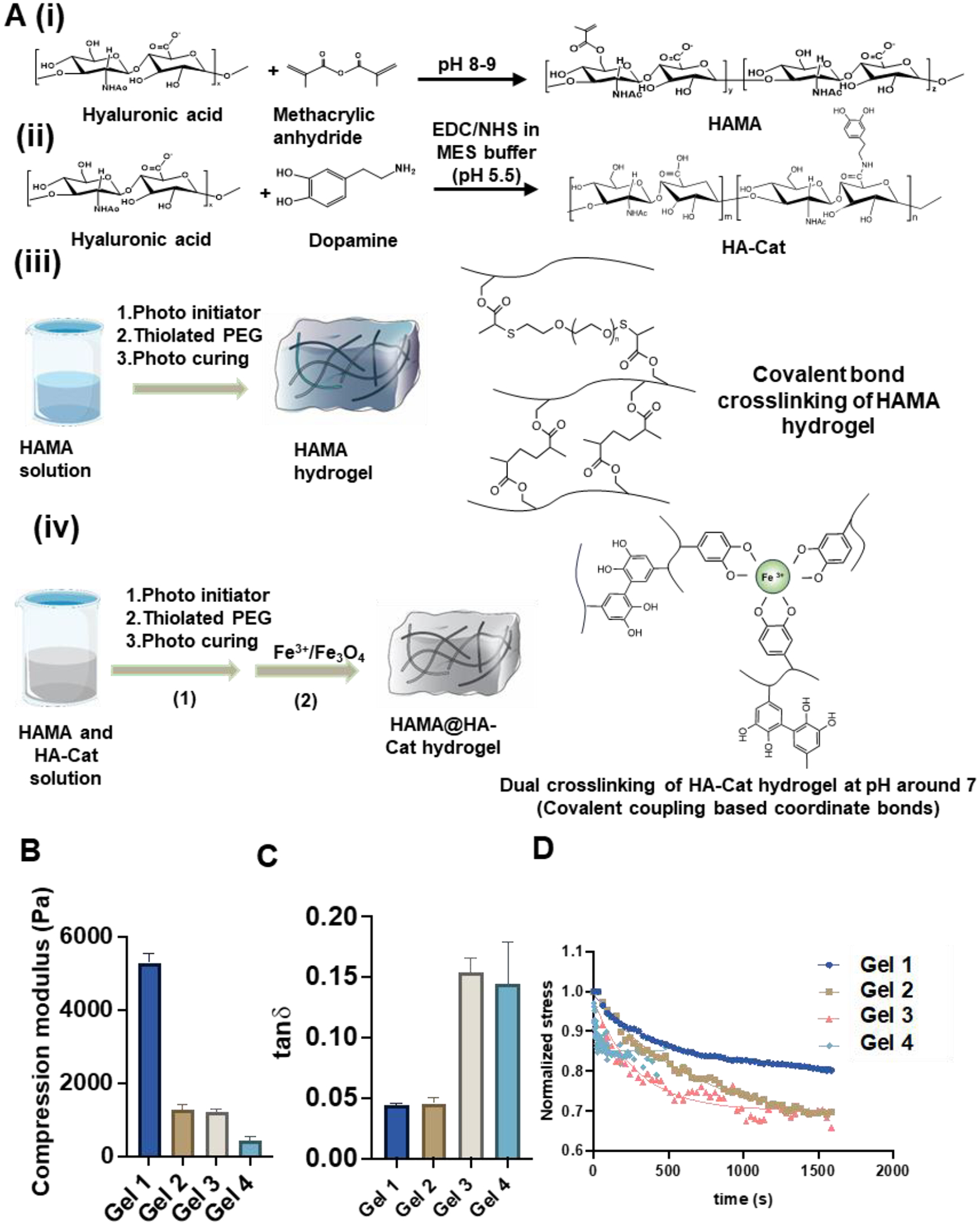
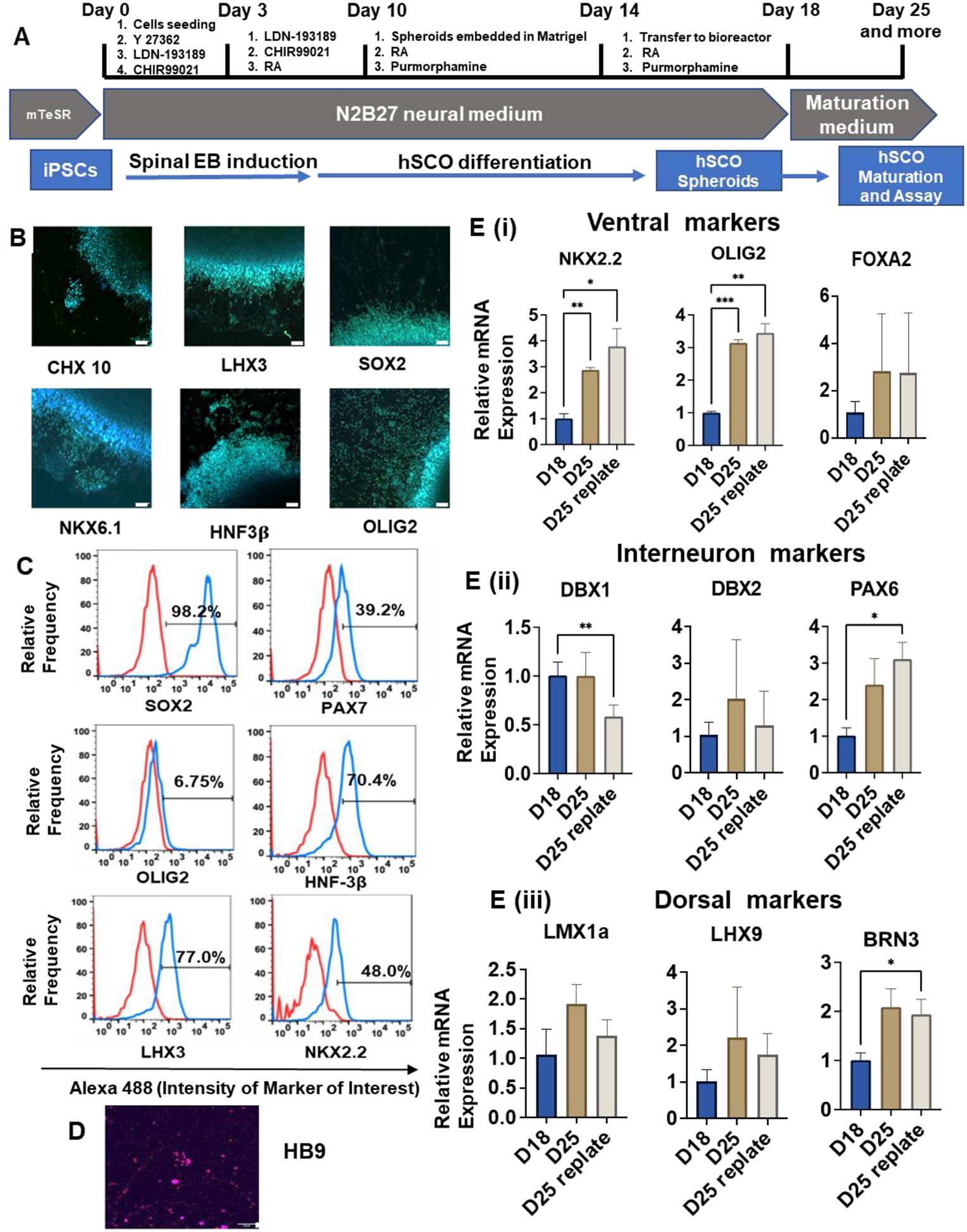
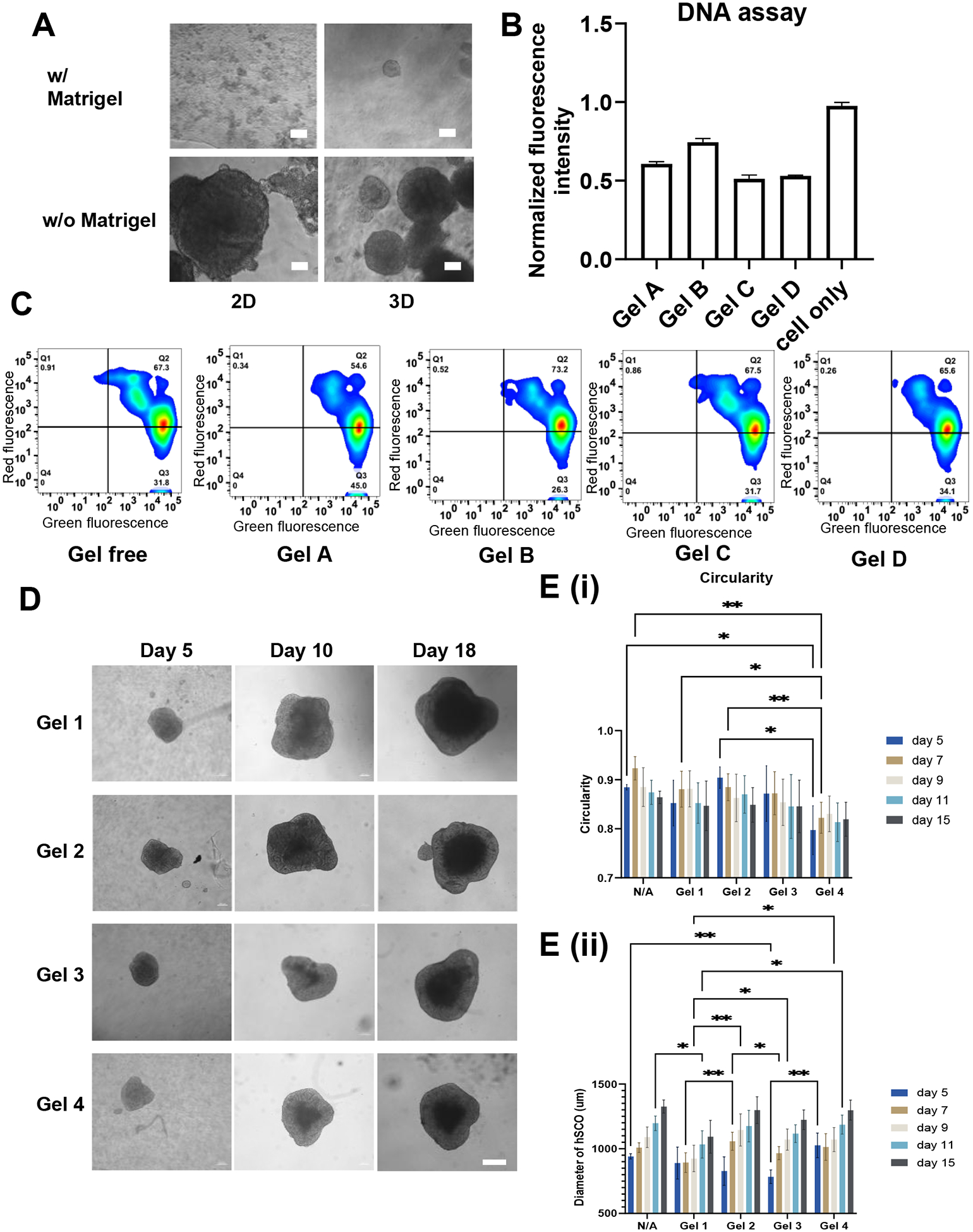
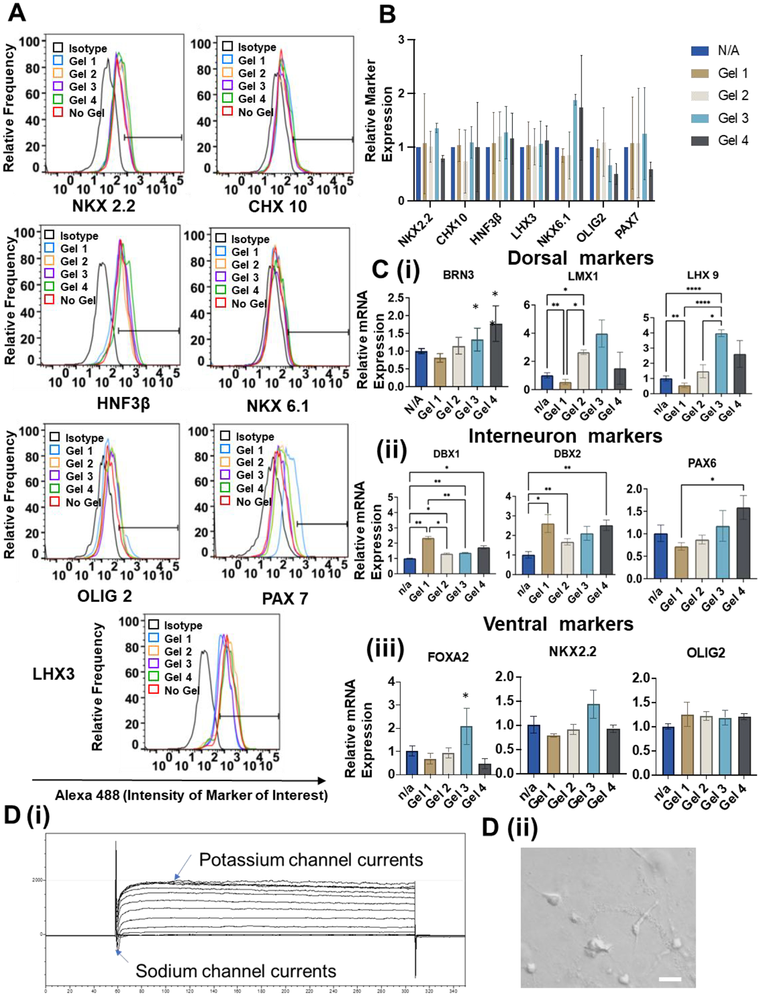
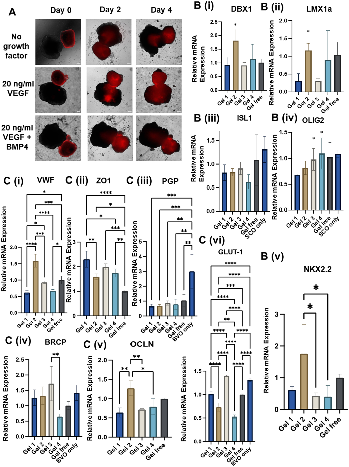
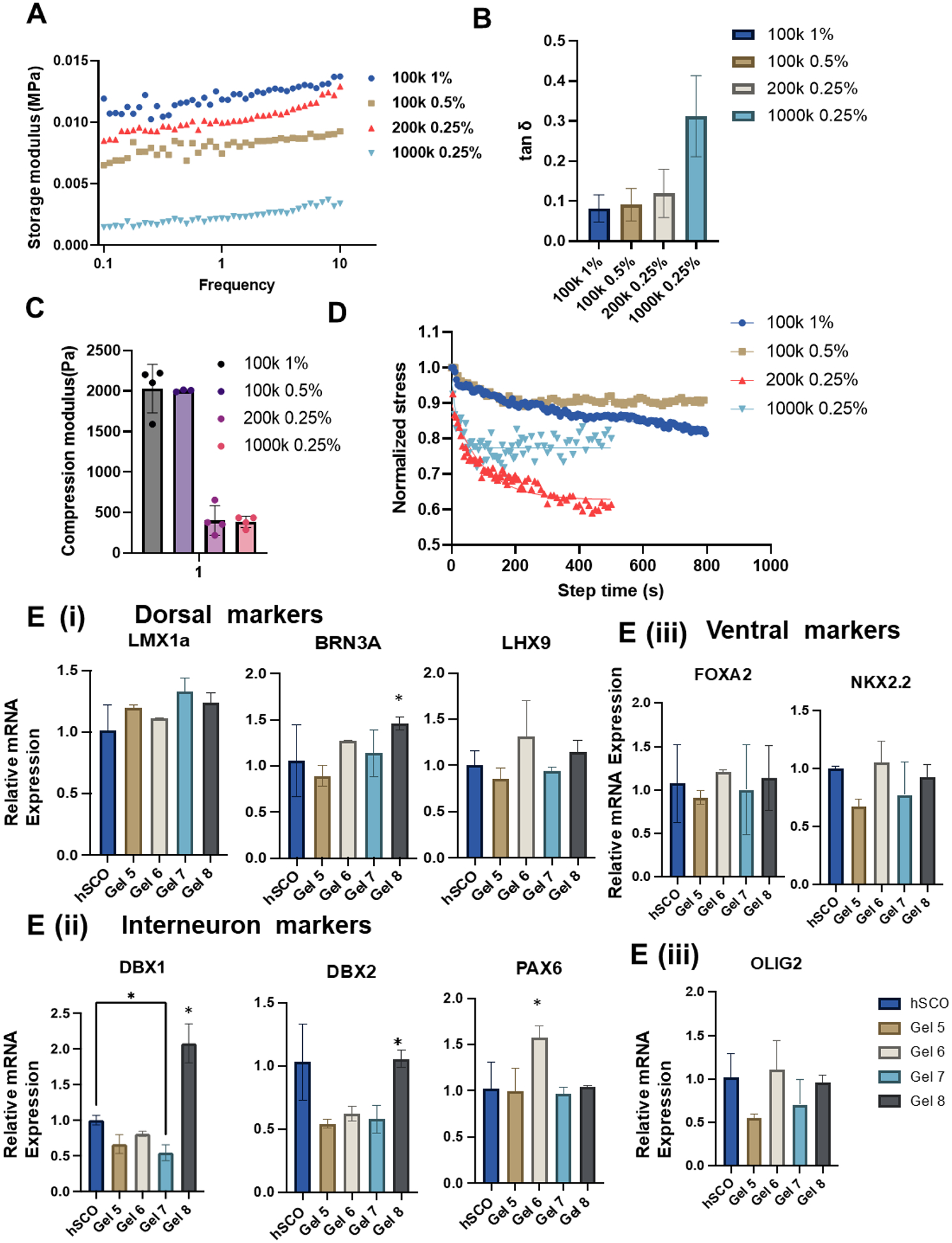
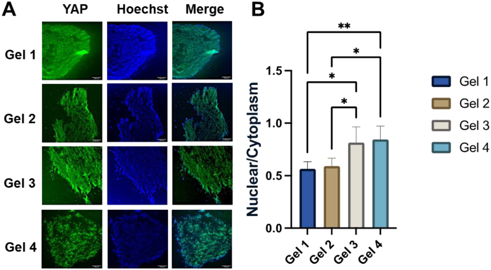
Recently, it has been recognized that natural extracellular matrix (ECM) and tissues are viscoelastic, while only elastic properties have been investigated in the past. How the viscoelastic matrix regulates stem cell patterning is critical for cell-ECM mechano-transduction. Here, this study fabricated different methacrylated hyaluronic acid (HA) hydrogels using covalent cross-linking, consisting of two gels with similar elasticity (stiffness) but different viscoelasticity, and two gels with similar viscoelasticity but different elasticity (stiffness). Meanwhile, a second set of dual network hydrogels are fabricated containing both covalent and coordinated cross-links. Human spinal cord organoid (hSCO) patterning in HA hydrogels and co-culture with isogenic human blood vessel organoids (hBVOs) are investigated. The viscoelastic hydrogels promote regional hSCO patterning compared to the elastic hydrogels. More viscoelastic hydrogels can promote dorsal marker expression, while softer hydrogels result in higher interneuron marker expression. The effects of viscoelastic properties of the hydrogels become more dominant than the stiffness effects in the co-culture of hSCOs and hBVOs. In addition, more viscoelastic hydrogels can lead to more Yes-associated protein nuclear translocation, revealing the mechanism of cell-ECM mechano-transduction. This research provides insights into viscoelastic behaviors of the hydrogels during human organoid patterning with ECM-mimicking in vitro microenvironments for applications in regenerative medicine.

Keywords: human pluripotent stem cells; hyaluronic acid hydrogels; spinal cord organoid patterning; vascularization, viscoelasticity.


Conflict of interest statement

Competing interests:

Authors declare that they have no competing interests.

Figures

Figure 1.
Figure 1.. HAMA synthesis and characterization.
(A) Schematic illustration of methods of HAMA and HA-Cat synthesis. (i-iii) Schematic process of fabrication and synthesis of (i) HAMA, (ii) HAMA@HA-Cat, and (iii) hydrogels. (iv) Schematic illustration of the process of Fe3+ curing HA-Cat hydrogels. (B) Quantification of the compression modulus, and (C) tanδ of the hydrogels by compression and rheological test. (n > 3 measurements per gel). (D) Stress relaxation test was applied to the 4 selected HAMA hydrogels and regression was performed by a modified Maxwell model to get stress relaxation time.
Figure 2.
Figure 2.. Ventral hSCO differentiation and characterization.
(A) Schematic illustration of ventral hSCO differentiation protocol. (B, C, D) Immunostaining and flow cytometry analysis for marker expression of hSCO differentiation. (B) and (D) were taken using confocal microscopy. Scale bar = 50 μm. (E) Quantitative RT-PCR for relative mRNA expression of various spinal cord markers after biochemical induction (n=3). (i) Ventral markers; (ii) Interneuron markers; (iii) Dorsal markers. * indicates p≤0.05, **: p≤0.01, ***: p≤0.001.
Figure 3.
Figure 3.. Biocompatibility of the HAMA hydrogels and morphogenesis of the organoids.
(A) hiPSC culture with HAMA and HAMA/Matrigel mixture for 7 days. Scale bar = 50 μm. (B) DNA assay and (C) Live/Dead flow cytometry analysis for determining proliferation rate and survival rate of hiPSCs cultured with different HAMA hydrogels, respectively. (D) Images of morphology of the organoids with different hydrogels over the time. Scale bar = 200 μm. (E) Quantification of diameter and circularity of hSCOs cultured in different HAMA hydrogels for morphogenesis. * indicates p≤0.05, **: p≤0.01, ***: p≤0.001.
Figure 4.
Figure 4.. Characterization for differentiation of hSCOs in different HAMA hydrogels.
(A) Flow cytometry analysis of expression of different ventral markers when generating hSCOs in different hydrogels. (B) Summary of 3 runs of flow cytometry analysis for identification ventral hSCO marker expression. (C) RT-PCR analysis of relative mRNA expression for different region-specific patterning markers during generation of hSCOs at day 35 (n=3). * indicates p≤0.05, **: p≤0.01, ***: p≤0.001. (D) (i) Electrophysiology to show sodium and potassium currents for the replated hSCOs at day 40. (ii) Morphology of outgrowth cells of the replated hSCOs for electrophysiology. Scale bar = 20 μm.
Figure 5.
Figure 5.. hSCO and hBVO coculturing for Blood-Spinal Cord Barrier (BSCB) generation.
(A) Morphology of the merging process of two types of organoids indicated by cell-tracker (red) hBVOs. (B, C) RT-PCR analysis for relative mRNA expression of ventral spinal cord genes, endothelial cells (EC), and blood-brain barrier (BBB) genes during hBVO and different hSCO coculturing. n=3, ns: p>0.05, * indicates p≤0.05, **: p≤0.01, ***: p≤0.001.
Figure 6.
Figure 6.. Fabrication and characterization of HAMA@HA-Cat hydrogels.
The dynamic hydrogels were fabricated to enhance the hydrogel properties and potential ability to regulate hSCO derivation. (A, B, C) Rheological test and compression test were performed to determine mechanical properties for the four new hydrogels (Gel 5–8 in sequence). (A) Storage modulus; (B) tanδ; (C) Compression modulus; (D) The viscoelasticity of the hydrogels was further determined by stress relaxation test. (E) RT-PCR analysis of relative mRNA expression for different region-specific patterning markers of hSCOs at day 35. n=3, * indicates p≤0.05.
Figure 7.
Figure 7.. Histological sections for YAP localization to reveal the mechanism of hydrogel effects on hSCO patterning.
(A) Images of YAP localization. Scale bar: 50 μm. (B) The quantitative measurements of nuclear to cytoplasmic YAP localization for different hydrogel conditions. * indicates p≤0.05, **: p≤0.01.

References

    1. D’Mello R, Dickenson AH, Spinal cord mechanisms of pain, British journal of anaesthesia 101(1) (2008) 8–16. - PubMed
    1. Revah O, Gore F, Kelley KW, Andersen J, Sakai N, Chen X, Li MY, Birey F, Yang X, Saw NL, Baker SW, Amin ND, Kulkarni S, Mudipalli R, Cui B, Nishino S, Grant GA, Knowles JK, Shamloo M, Huguenard JR, Deisseroth K, Pasca SP, Maturation and circuit integration of transplanted human cortical organoids, Nature 610(7931) (2022) 319–326. - PMC - PubMed
    1. Du Z-W, Chen H, Liu H, Lu J, Qian K, Huang C-L, Zhong X, Fan F, Zhang S-C, Generation and expansion of highly pure motor neuron progenitors from human pluripotent stem cells, Nature communications 6(1) (2015) 1–9. - PMC - PubMed
    1. Yan Y, Li X, Gao Y, Mathivanan S, Kong L, Tao Y, Dong Y, Li X, Bhattacharyya A, Zhao X, Zhang SC, 3D bioprinting of human neural tissues with functional connectivity, Cell Stem Cell 31(2) (2024) 260–274 e7. - PMC - PubMed
    1. Ogura T, Sakaguchi H, Miyamoto S, Takahashi J, Three-dimensional induction of dorsal, intermediate and ventral spinal cord tissues from human pluripotent stem cells, Development 145(16) (2018). - PMC - PubMed

LinkOut - more resources