

Optical imaging for screening and early cancer diagnosis in low-resource settings
- PMID: 39301200
- PMCID: PMC11412616
- DOI: 10.1038/s44222-023-00135-4
Optical imaging for screening and early cancer diagnosis in low-resource settings
Abstract
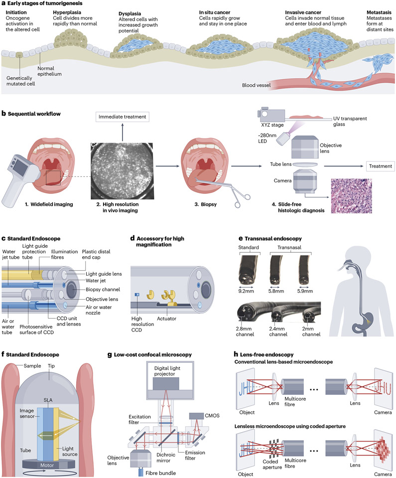
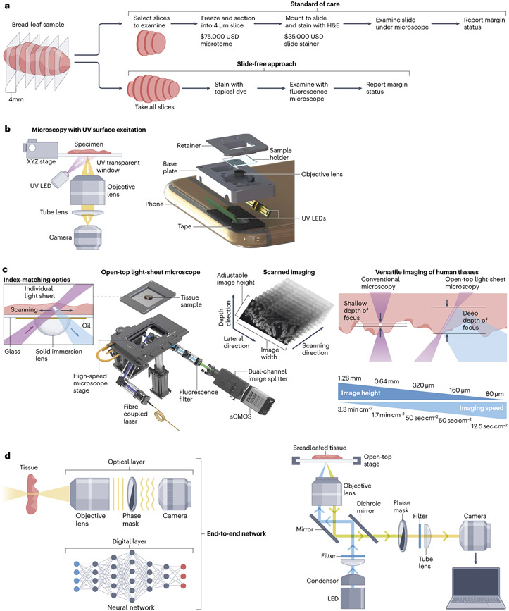
Low-cost optical imaging technologies have the potential to reduce inequalities in healthcare by improving the detection of pre-cancer or early cancer and enabling more effective and less invasive treatment. In this Review, we summarise technologies for in vivo widefield, multi-spectral, endoscopic, and high-resolution optical imaging that could offer affordable approaches to improve cancer screening and early detection at the point-of-care. Additionally, we discuss approaches to slide-free microscopy, including confocal imaging, lightsheet microscopy, and phase modulation techniques that can reduce the infrastructure and expertise needed for definitive cancer diagnosis. We also evaluate how machine learning-based algorithms can improve the accuracy and accessibility of optical imaging systems and provide real-time image analysis. To achieve the potential of optical technologies, developers must ensure that devices are easy to use; the optical technologies must be evaluated in multi-institutional, prospective clinical tests in the intended setting; and the barriers to commercial scale-up in under-resourced markets must be overcome. Therefore, test developers should view the production of simple and effective diagnostic tools that are accessible and affordable for all countries and settings as a central goal of their profession.
Figures
Similar articles
-
The future of Cochrane Neonatal.Early Hum Dev. 2020 Nov;150:105191. doi: 10.1016/j.earlhumdev.2020.105191. Epub 2020 Sep 12. Early Hum Dev. 2020. PMID: 33036834
-
Optical endomicroscopy and the road to real-time, in vivo pathology: present and future.Diagn Pathol. 2012 Aug 13;7:98. doi: 10.1186/1746-1596-7-98. Diagn Pathol. 2012. PMID: 22889003 Free PMC article. Review.
-
Emerging roles for multimodal optical imaging in early cancer detection: a global challenge.Technol Cancer Res Treat. 2010 Apr;9(2):211-7. doi: 10.1177/153303461000900210. Technol Cancer Res Treat. 2010. PMID: 20218743 Free PMC article. Review.
-
Strategies for Early Keratoconus Diagnosis: A Narrative Review of Evaluating Affordable and Effective Detection Techniques.J Clin Med. 2025 Jan 13;14(2):460. doi: 10.3390/jcm14020460. J Clin Med. 2025. PMID: 39860468 Free PMC article. Review.
-
New optical imaging technologies for bladder cancer: considerations and perspectives.J Urol. 2012 Aug;188(2):361-8. doi: 10.1016/j.juro.2012.03.127. Epub 2012 Jun 13. J Urol. 2012. PMID: 22698620 Free PMC article. Review.
Cited by
-
Lightweight Low-Rank Adaptation Vision Transformer Framework for Cervical Cancer Detection and Cervix Type Classification.Bioengineering (Basel). 2024 May 8;11(5):468. doi: 10.3390/bioengineering11050468. Bioengineering (Basel). 2024. PMID: 38790335 Free PMC article.
-
Integrating artificial intelligence with smartphone-based imaging for cancer detection in vivo.Biosens Bioelectron. 2025 Mar 1;271:116982. doi: 10.1016/j.bios.2024.116982. Epub 2024 Nov 21. Biosens Bioelectron. 2025. PMID: 39616900 Review.
-
Nanophotonic-enhanced photoacoustic imaging for brain tumor detection.J Nanobiotechnology. 2025 Mar 5;23(1):170. doi: 10.1186/s12951-025-03204-5. J Nanobiotechnology. 2025. PMID: 40045308 Free PMC article. Review.
-
Emerging Trends in Point-of-Care Technology Development for Oncology in Low- and Middle-Income Countries.JCO Glob Oncol. 2025 Jun;11:e2500142. doi: 10.1200/GO-25-00142. Epub 2025 Jun 18. JCO Glob Oncol. 2025. PMID: 40532137 Free PMC article. Review.
-
Nano optical (bio)sensing platforms for early detection of pediatric cancer.Mikrochim Acta. 2025 Jun 5;192(7):406. doi: 10.1007/s00604-025-07246-2. Mikrochim Acta. 2025. PMID: 40471347 Review.
References
-
- Wild CP, Weiderpass E, Stewart BW (Eds). World Cancer Report: Cancer Research for Cancer Prevention. (International Agency for Research on Cancer, 2020).
-
- American Association for Cancer Research. Cancer Disparities Progress Report. (2022).
-
- Mitchell E et al. Cancer healthcare disparities among African Americans in the United States. J Natl Med Assoc 114, 236–250 (2022). - PubMed
Grants and funding
LinkOut - more resources
Full Text Sources