

The frontal association area: exercise-induced brain plasticity in children and adolescents and implications for cognitive intervention practice
- PMID: 39301538
- PMCID: PMC11410640
- DOI: 10.3389/fnhum.2024.1418803
The frontal association area: exercise-induced brain plasticity in children and adolescents and implications for cognitive intervention practice
Abstract
Objective: Explore the plasticity of the frontal associative areas in children and adolescents induced by exercise and potential moderating variables.
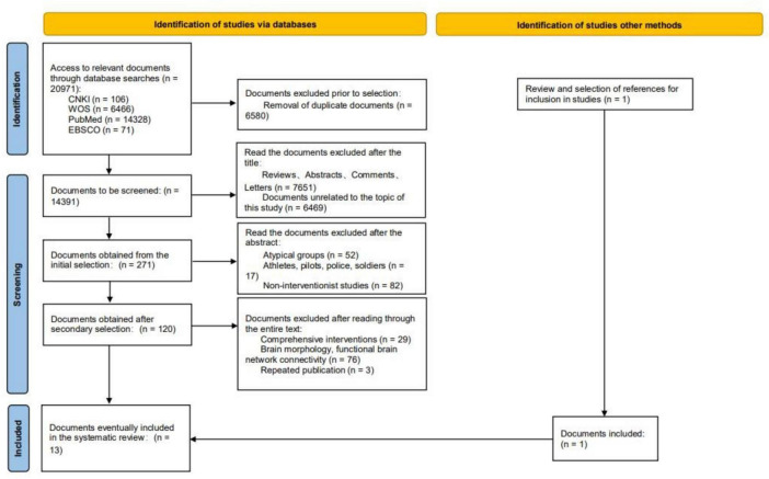
Methods: Computer searches of CNKI, WOS, PubMed and EBSCO databases were conducted, and statistical analyses were performed based on SPSS 25.0, Stata 12.0 and Ginger ALE 2.3 software after literature screening, data extraction and quality assessment were performed independently by two researchers.
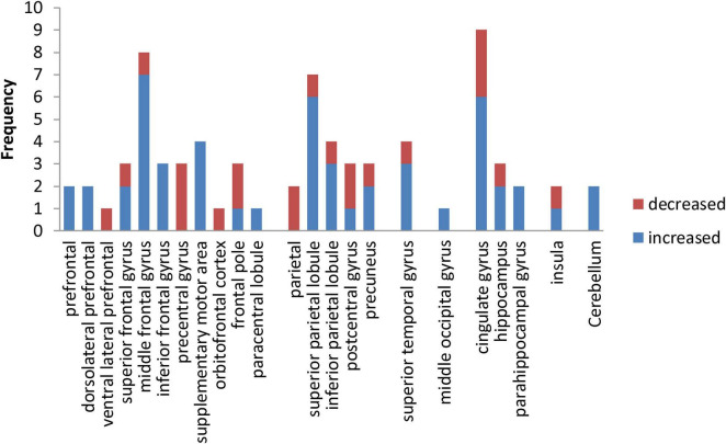
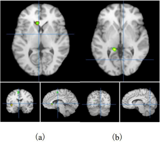
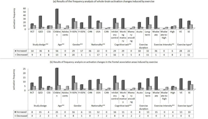
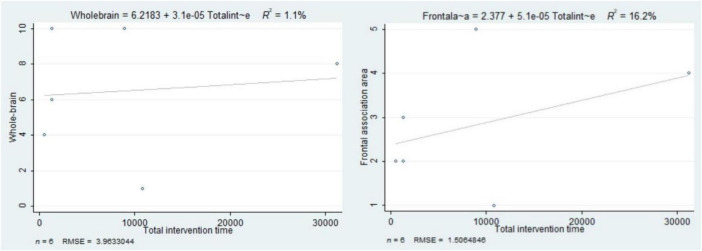
Results: A total of 13 articles, including 425 participants aged 8.9∼16.8 years, were included. Frequency analysis revealed that exercise induced enhanced activation in frontal, parietal, occipital, limbic system and cerebellum (P < 0.01). Activation Likelihood Estimation (ALE) meta-analysis revealed that exercise altered the activation status of the frontal association (medial frontal gyrus, middle frontal gyrus, inferior frontal gyrus and precentral gyrus), cuneus, lingual gyrus, cingulate gyrus, parahippocampal gyrus, caudate nucleus and cerebellar apex, with the volume of activation in the frontal association accounting for 61.81% of the total activation cluster volume and an enhanced activation effect. Additionally, the study design, age, gender, nationality, cognitive tasks, as well as exercise intensity, intervention time, and type of exercise may be potential moderating variables. Particularly, sustained exercise induced a decrease in activation in the left parahippocampal gyrus, culmen, and lingual gyrus, while variable exercise induced an increase in activation in the left middle frontal gyrus.
Conclusion: Exercise-induced activation increase in the frontal associative areas of children and adolescents is dominant, especially longer periods of moderate-intensity variable exercise can induce more brain region activation. However, some of the included studies are cross-sectional, and the accuracy of the results still requires further verification.
Systematic review registration: https://www.crd.york.ac.uk/prospero/, identifier PROSPERO, CRD42022348781.
Keywords: ALE meta-analysis; brain; exercise; fMRI; frontal association area; neuroplasticity.
Copyright © 2024 Zhang, Shi, Zhang, Li and Feng.
Conflict of interest statement
The authors declare that the research was conducted in the absence of any commercial or financial relationships that could be construed as a potential conflict of interest.
Figures
Similar articles
-
Neural effects of acupuncture on stroke patients with motor dysfunction: an activation likelihood estimation meta-analysis.Front Neurol. 2024 Sep 25;15:1453935. doi: 10.3389/fneur.2024.1453935. eCollection 2024. Front Neurol. 2024. PMID: 39385820 Free PMC article.
-
Shared and Distinct Neural Bases of Large- and Small-Scale Spatial Ability: A Coordinate-Based Activation Likelihood Estimation Meta-Analysis.Front Neurosci. 2019 Jan 10;12:1021. doi: 10.3389/fnins.2018.01021. eCollection 2018. Front Neurosci. 2019. PMID: 30686987 Free PMC article.
-
Effects of open-skill exercise on executive functions in children and adolescents: a systematic review and meta-analysis.Front Hum Neurosci. 2025 Feb 4;18:1495371. doi: 10.3389/fnhum.2024.1495371. eCollection 2024. Front Hum Neurosci. 2025. PMID: 39967690 Free PMC article.
-
Activation of brain regions using task-state FMRI in patients with mild traumatic brain injury: a meta-analysis.Int J Clin Exp Pathol. 2020 Dec 1;13(12):2918-2926. eCollection 2020. Int J Clin Exp Pathol. 2020. PMID: 33425093 Free PMC article. Review.
-
Effect of acupuncture on brain functional networks in patients with mild cognitive impairment: an activation likelihood estimation meta-analysis.Acupunct Med. 2023 Oct;41(5):259-267. doi: 10.1177/09645284221146199. Epub 2023 Feb 15. Acupunct Med. 2023. PMID: 36790017
Cited by
-
The effect of physical activity on resilience of Chinese children: the chain mediating effect of executive function and emotional regulation.BMC Pediatr. 2025 Jul 22;25(1):563. doi: 10.1186/s12887-025-05883-3. BMC Pediatr. 2025. PMID: 40696298 Free PMC article.
-
Exercise prescription to improve inhibitory control in children and adolescents with ADHD: a network meta-analysis.Front Psychiatry. 2025 Jun 30;16:1601765. doi: 10.3389/fpsyt.2025.1601765. eCollection 2025. Front Psychiatry. 2025. PMID: 40661882 Free PMC article.
-
Longitudinal Reciprocal Effects of Physical Exercise, Executive Function, and Subjective Well-Being: A Three-Wave Random-Intercept Cross-Lagged Panel Model in Chinese Minority College Students.Behav Sci (Basel). 2025 Jun 26;15(7):865. doi: 10.3390/bs15070865. Behav Sci (Basel). 2025. PMID: 40723649 Free PMC article.
References
-
- Bohbot V. D., Allen J. J., Dagher A., Dumoulin S. O., Evans A. C., Petrides M., et al. (2015). Role of the parahippocampal cortex in memory for the configuration but not the identity of objects: Converging evidence from patients with selective thermal lesions and fMRI. Front. Hum. Neurosci. 9:431. 10.3389/fnhum.2015 - DOI - PMC - PubMed
-
- Cai Q. T. (2014). National core literacy. Taipei: Higher Education and Culture Business Ltd, 162.
Publication types
LinkOut - more resources
Full Text Sources
Miscellaneous