. 2025 Mar 6;148(3):1001-1014.
doi: 10.1093/brain/awae302.

A role for leucine-rich, glioma inactivated 1 in regulating pain sensitivity

Affiliations

A role for leucine-rich, glioma inactivated 1 in regulating pain sensitivity

Adham Farah et al. Brain. .

Abstract

Neuronal hyperexcitability is a key driver of persistent pain states, including neuropathic pain. Leucine-rich, glioma inactivated 1 (LGI1) is a secreted protein known to regulate excitability within the nervous system and is the target of autoantibodies from neuropathic pain patients. Therapies that block or reduce antibody levels are effective at relieving pain in these patients, suggesting that LGI1 has an important role in clinical pain. Here we have investigated the role of LGI1 in regulating neuronal excitability and pain-related sensitivity by studying the consequences of genetic ablation in specific neuron populations using transgenic mouse models. LGI1 has been well studied at the level of the brain, but its actions in the spinal cord and peripheral nervous system are poorly understood. We show that LGI1 is highly expressed in dorsal root ganglion (DRG) and spinal cord dorsal horn neurons in both mouse and human. Using transgenic mouse models, we genetically ablated LGI1, either specifically in nociceptors (LGI1fl/Nav1.8+) or in both DRG and spinal neurons (LGI1fl/Hoxb8+). On acute pain assays, we found that loss of LGI1 resulted in mild thermal and mechanical pain-related hypersensitivity when compared with littermate controls. In LGI1fl/Hoxb8+ mice, we found loss of Kv1 currents and hyperexcitability of DRG neurons. LGI1fl/Hoxb8+ mice displayed a significant increase in nocifensive behaviours in the second phase of the formalin test (not observed in LGI1fl/Nav1.8+ mice), and extracellular recordings in LGI1fl/Hoxb8+ mice revealed hyperexcitability in spinal dorsal horn neurons, including enhanced wind-up. Using the spared nerve injury model, we found that LGI1 expression was dysregulated in the spinal cord. LGI1fl/Nav1.8+ mice showed no differences in nerve injury-induced mechanical hypersensitivity, brush-evoked allodynia or spontaneous pain behaviour compared with controls. However, LGI1fl/Hoxb8+ mice showed a significant exacerbation of mechanical hypersensitivity and allodynia. Our findings point to effects of LGI1 at the level of both the DRG and the spinal cord, including an important impact of spinal LGI1 on pathological pain. Overall, we find a novel role for LGI1 with relevance to clinical pain.

Keywords: hyperexcitability; leucine-rich, glioma inactivated 1 (LGI1); mechanical pain hypersensitivity; neuropathic pain; wind-up.


Conflict of interest statement

No competing interests.

Figures

Figure 1
Figure 1
LGI1  expression in dorsal root ganglion (DRG) and spinal cord. (A and B) Representative images of mouse L4 DRG showing in situ hybridization (ISH) for LGI1 mRNA (red) in CGRP+ (green), NF200+ (blue) (A) and IB4+ neurons (B); scale bar = 50 µm. (C) No ISH signal is observed in DRG neurons when using a negative control probe. (D) Quantification of LGI1 mRNA signal intensity in DRG neurons (n = 3 or 4 mice). (E) Representative images of mouse lumbar spinal cord showing ISH for LGI1 mRNA (red), with Neurotrace (blue) to mark neurons and CGRP (green) to mark lamina IIo; scale bar = 100 µm. (F) Representative images of mouse lumbar spinal cord showing ISH for LGI1 mRNA (red), with Neurotrace to mark all neurons (blue) and Pax-2 (green) to mark inhibitory neurons; scale bar = 100 µm. (G) Quantification of LGI1 mRNA signal intensity in mouse lumbar spinal cord (n = 4 mice). All data are shown as the mean ± standard error of the mean (SEM).
Figure 2
Figure 2
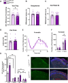
Conditional ablation of LGI1 increases pain-related behaviour in mice. (A) Using Von Frey hair application, LGI1fl/Hoxb8+ mice (n = 31) displayed mechanical pain-related hypersensitivity when compared with littermate controls (n = 25). (B and C) LGI1fl/Hoxb8+ mice did not display thermal hypersensitivity as measured by the Hargreaves test (B) and/or a hot plate set at 53°C (C) (LGI1fl/Hoxb8+  n = 22; LGI1fl/Hoxb8, n = 21). (D) Withdrawal latency to pin prick application was not altered in LGI1fl/Hoxb8+ mice (n = 15) versus controls (LGI1fl/Hoxb8, n = 16). (E) LGI1fl/Hoxb8+ mice (n = 14) display increased nocifensive behaviour during the second phase of the formalin test when compared with littermate controls (n = 10). (F) Total duration of nocifensive behaviour during both the first phase (0–15 min) and the second phase (15–60 min) of the formalin test. (G) The number of c-Fos+ neurons is increased in the ipsilateral superficial dorsal horn (laminae I + II, S. IPSI) and deeper laminae (III + IV, D. IPSI) of the spinal cord in LGI1fl/Hoxb8+ mice 2 h after formalin injection (n = 5) compared with littermate controls (n = 4). An increase in ipsilateral c-Fos expression is seen for both genotypes when compared with contralateral (CONTRA) spinal cord. (H) Representative images of c-Fos expression (green) in the ipsilateral spinal cord dorsal horn of LGI1fl/Hoxb8 and LGI1fl/Hoxb8+ mice following formalin injection. Neurons are marked with Neurotrace (red), with IB4 shown in blue; scale bar = 100 µm. Data are shown as the mean ± SEM; *P < 0.05 and **P < 0.01 versus LGI1fl/Hoxb8; #P < 0.05 and ##P < 0.01 versus contralateral. ns = not significant.
Figure 3
Figure 3
Dorsal root ganglion (DRG) neuron excitability in LGI1fl/Hoxb8+ mice. (A and B) Calcium imaging data for DRG neurons cultured from LGI1fl/Hoxb8+ and LGI1fl/Hoxb8 mice (n = 4 mice, data taken from ∼500 cells per genotype) during the initial untreated period [spontaneous (S) activity] and in response to both ATP (10 μM) and capsaicin (CAP, 1 μM). No difference was seen between genotypes for both the percentage of DRG neurons responding (A) and the amplitude of their response (B, left). Neurons were identified by their response to 50 mM KCl. (B, right) Example traces of calcium transients. (C) Whole-cell patch-clamp electrophysiology revealed no difference in the rheobase of both small (<25 μm in diameter, n = 46 or 47 cells taken from four mice) or medium-sized (25–35 μm, n = 23 taken from four mice) DRG neurons between genotypes. (D and E) Action potential firing in response to a range of prolonged current injections in both small cells (D) and medium cells (E). (D) Small DRG neurons from LGI1fl/Hoxb8+ mice displayed significantly increased firing in comparison to controls. Representative traces are also shown. (F) Example traces of outward current in small DRG neurons (<25 µm in diameter) from both LGI1fl/Hoxb8+ and LGI1fl/Hoxb8 mice evoked by depolarizing pulses. (G) IKD (slowly-inactivating potassium current) was measured before and after 100 nM dendrotoxin (DTX). Current–voltage relationships for IKD pre-DTX (total IKD) were significantly reduced in LGI1fl/Hoxb8+ neurons (n = 14) compared with controls (n = 14). (H) DTX-sensitive currents (total IKDIKD + DTX) were significantly reduced in small-diameter neurons from LGI1fl/Hoxb8+ mice. All data are shown as the mean ± SEM. (D) **P < 0.01 versus LGI1fl/Hoxb8 neurons. (G) *P < 0.05 versus LGI1fl/Hoxb8+.
Figure 4
Figure 4
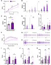
LGI1 ablation leads to spinal hyperexcitability in LGI1fl/Hoxb8+ mice. (A) Responses to punctate mechanical stimulation are enhanced in wide dynamic range (WDR) neurons from LGI1fl/Hoxb8+ mice compared with controls. (B) Representative histogram traces of single-unit responses to mechanical stimulation. (C) Responses to dynamic brushing are increased in WDR neurons from LGI1fl/Hoxb8+ mice compared with controls. (D) Representative histogram traces of single-unit responses to brush stimulation. (E) No differences were found between genotypes for electrical activation thresholds of WDR neurons in response to both A- and C-fibre input. (F) Wind-up curve plotted as total evoked spikes per stimulus. (G and H) Representative neurogram traces for LGI1fl/Hoxb8 (G) and LGI1fl/Hoxb8+ (H) mice of single-unit responses, separated by latency for the first and last stimulus. (I) Total non-potentiated response (NPR) and wind-up (WU) are significantly increased in LGI1fl/Hoxb8+ mice compared with controls. (J) Neuronal events evoked by the first electrical stimulus separated by latency. Spikes during A- and C-fibre latencies and post discharge (PD) are increased in LGI1fl/Hoxb8+ mice. (K) Total neuronal events evoked by 16 electrical stimuli separated by latency. Spikes during A- and C-fibre latencies and during post discharge are increased in LGI1fl/Hoxb8+ mice. All data are shown as the mean ± SEM; *P < 0.05, **P < 0.01 and ***P < 0.001 versus LGI1fl/Hoxb8 mice (LGI1fl/Hoxb8, n = 13 mice; LGI1fl/Hoxb8+, n = 13 mice).
Figure 5
Figure 5
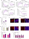
Neuropathic pain is exacerbated in LGI1fl/Hoxb8+ mice. (A) Both LGI1fl/Hoxb8 (n = 14) and LGI1fl/Hoxb8+ (n = 17) mice developed ipsilateral mechanical hypersensitivity following nerve injury, as measured by Von Frey hair application. Ipsilateral mechanical hypersensitivity was significantly increased in LGI1fl/Hoxb8+ mice compared with controls. (B) Percentage increase in mechanical sensitivity, normalized to baseline, as measured by Von Frey hair application. (C) Both LGI1fl/Hoxb8 and LGI1fl/Hoxb8+ mice developed ipsilateral mechanical allodynia following nerve injury, as measured by brush application. (AC) LGI1fl/Hoxb8+ mice also developed significant contralateral pain hypersensitivity versus controls. (D) All mice developed cold hypersensitivity following nerve injury as measured by a decrease in the time spent at 16°C, but no differences were seen between genotypes. (E) Conditioned place preference test showing that on average mice from both genotypes developed a preference for the gabapentin paired chamber, indicating the development of spontaneous pain. However, no differences were seen between LGI1fl/Hoxb8 (n = 6) and LGI1fl/Hoxb8+ (n = 8) mice. (F and G) Representative images of L4 mouse dorsal root ganglia (DRG) showing in situ hybridization (ISH) for LGI1 mRNA (red) in naïve wild-type (WT) mice (F) or 7 days after nerve injury (G). Neurotrace (blue) was used to mark all neurons; ATF3 (green) shows injured neurons. (H) Quantification of LGI1 mRNA signal in DRG neurons before (No SNI, n = 4) and 7 days after injury (n = 3), including both injured (ATF3+) and uninjured (ATF3−) neurons; scale bar = 50 µm. (I and J) Representative images of mouse L4 lumbar spinal cord showing ISH for LGI1 mRNA (red) in naïve WT mice (I) or 7 days after nerve injury (J). Neurotrace (blue) was used to mark all neurons. (K) Quantification of LGI1 mRNA signal in L4 spinal cord. An increase in expression was seen in the ipsilateral spinal cord 7 days after injury (n = 3) compared with naïve controls (n = 4). No significant increase was seen in the contralateral cord after injury. (L) Quantification of LGI1 mRNA signal in L4 spinal cord before and 7 days after injury separated by laminae. All data are shown as the mean ± SEM. (AC) *P < 0.05, **P < 0.01 and ***P < 0.001 versus LGI1fl/Hoxb8 at specific time points; ###P < 0.001 versus LGI1fl/Hoxb8. (D) *P < 0.05, ***P < 0.001, +P < 0.05 and +++P < 0.001 versus baseline (BL), ns (not significant) between genotypes. (E) ***P < 0.001 versus saline condition for all mice, ns between genotypes . (K) *P < 0.05 versus No spared nerve injury (SNI).

References

    1. Van Hecke O, Austin SK, Khan RA, Smith BH, Torrance N. Neuropathic pain in the general population: A systematic review of epidemiological studies. Pain. 2014;155:654–662. - PubMed
    1. Baskozos G, Hébert HL, Pascal MMV, et al. Epidemiology of neuropathic pain: An analysis of prevalence and associated factors in UK biobank. Pain Rep. 2023;8:e1066. - PMC - PubMed
    1. Fornasari D. Pharmacotherapy for neuropathic pain: A review. Pain Ther. 2017;6(Suppl 1):25–33. - PMC - PubMed
    1. Serra J, Bostock H, Solà R, et al. Microneurographic identification of spontaneous activity in C-nociceptors in neuropathic pain states in humans and rats. Pain. 2012;153:42–55. - PubMed
    1. Djouhri L, Zeidan A, Alzoghaibi M, Al Otaibi MF, Abd El-Aleem SA. L5 spinal nerve axotomy induces distinct electrophysiological changes in axotomized L5- and adjacent L4-dorsal root ganglion neurons in rats in vivo. J Neurotrauma. 2021;38:330–341. - PubMed