Review
. 2024 Dec;13(32):e2402001.
doi: 10.1002/adhm.202402001. Epub 2024 Sep 20.

Antimicrobial Biomaterials Based on Physical and Physicochemical Action

Affiliations
Review

Antimicrobial Biomaterials Based on Physical and Physicochemical Action

Adrian G Nowotnick et al. Adv Healthc Mater. 2024 Dec.

Abstract

Developing effective antimicrobial biomaterials is a relevant and fast-growing field in advanced healthcare materials. Several well-known (e.g., traditional antibiotics, silver, copper etc.) and newer (e.g., nanostructured, chemical, biomimetic etc.) approaches have been researched and developed in recent years and valuable knowledge has been gained. However, biomaterials associated infections (BAIs) remain a largely unsolved problem and breakthroughs in this area are sparse. Hence, novel high risk and potential high gain approaches are needed to address the important challenge of BAIs. Antibiotic free antimicrobial biomaterials that are largely based on physical action are promising, since they reduce the risk of antibiotic resistance and tolerance. Here, selected examples are reviewed such antimicrobial biomaterials, namely switchable, protein-based, carbon-based and bioactive glass, considering microbiological aspects of BAIs. The review shows that antimicrobial biomaterials mainly based on physical action are powerful tools to control microbial growth at biomaterials interfaces. These biomaterials have major clinical and application potential for future antimicrobial healthcare materials without promoting microbial tolerance. It also shows that the antimicrobial action of these materials is based on different complex processes and mechanisms, often on the nanoscale. The review concludes with an outlook and highlights current important research questions in antimicrobial biomaterials.

Keywords: antimicrobials; bioglass; biomaterial associated infections; graphene; physical actions; proteins; switching.


Conflict of interest statement

The authors declare no conflict of interest.

Figures
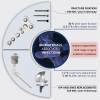
Figure 1
Figure 1
Fracture fixation elements, hip and knee replacements. These biomaterials applications are frequently connected to biomaterials associated infections (BAIs). Titanium and its alloys as well as ultra‐high molecular weight poly(ethylene) (UHMWPE) (knee) are typically used materials for these implants.
Figure 2
Figure 2
Illustrates the prevalence of key pathogenic species in orthopedic clinical isolates associated with implant‐related infections. CNS: coagulase‐negative staphylococci. Adapted with permission.[ 29 ] 2006, Elsevier.
Figure 3
Figure 3
Potential dependent states of PSBEDOT and the respective fluorescence microscopy images of microbial adhesion. Reproduced with permission.[ 57 ] 2016, Cao et al. (https://doi.org/10.1039/C5SC03887A) under Creative Commons license CC BY 3.0.
Figure 4
Figure 4
Schematic illustration of thermally induced hydration transition of nanopatterned PNIPAAm resulting in the release of a dead microbe (E. coli). Reproduced with permission.[ 63 ] 2014, RSC Publishing.
Figure 5
Figure 5
A hydrogel composed of the PCBSA polymer demonstrates the capability to maintain a microbe‐free surface and hinder microbial proliferation throughout its bulk. Reproduced with permission.[ 65 ] 2012, Elsevier.
Figure 6
Figure 6
Antimicrobial activity of glass surfaces without and with lysozyme over time. Black, blue, red lines correspond to surfaces functionalized with 1) biotin‐streptavidin, 2) lysozyme and 3) biotin‐streptavidin followed by lysozyme, respectively. Three challenges were performed. In contrast to lysozyme‐unsupported surfaces, the antibacterial activity of surfaces supported with lysozyme is preserved over consecutive challenges. Reproduced with permission.[ 96 ] 2021, Elsevier.
Figure 7
Figure 7
Impact of FN on S. aureus, S. epidermidis HAM892, and S. epidermidis ATCC35984 adhesion. Total number of microbes (n60) after 1 h of deposition on nonirradiated (control, C) and irradiated (UV) TiAl6V4 substrata in the absence and presence of FN coatings. Adapted with permission.[ 99 ] 2013, John Wiley and Sons.
Figure 8
Figure 8
Interaction of E. coli with fibrinogen nanofibers. A) Spectrophotometric tests show lower bacteria counts on nanofibrous fibrinogen than control agar, confirmed by statistical analysis. B) Top view images reveal dense E. coli on fibrinogen scaffolds. C) Top view images reveal fewer E. coli on fibrinogen scaffolds. D) SEM analysis shows minimal E. coli on fibrinogen scaffolds, with cell wall disruption indicated by white arrows. Reproduced with permission.[ 83 ] 2021, Elsevier.
Figure 9
Figure 9
a) Scanning electron microscopy (SEM) images of E. coli cells interacting with MWNTs. a) Cells incubated with MWNTs for 60 min. b) Cells incubated with SWNTs for 60 min. The bars in both images represent 2 µm. c) Concentrations of plasmid DNA and RNA in solution in the presence and absence of MWNTs. Reproduced with permission.[ 125 ] 2008, American Chemical Society.
Figure 10
Figure 10
Schematic representation of the interaction between graphene oxide (GO) and pathogens and the toxicity mechanisms of antibacterial activity of GO against bacterial phytopathogens and fungal spores. The bacterial cells and fungal spores intertwined with a wide range of aggregated GO sheets. This resulted in local perturbation of the cell membrane, which induced loss of bacterial membrane potential and leakage of electrolytes of fungal spores and caused the lysis and death of pathogens. Reproduced with permission.[ 145 ] 2014, RSC Publishing.
Figure 11
Figure 11
a) Schematic representation of one of the common proposed models for the antibacterial mechanism of graphene (G)/GO. b) Experimentally observed antibacterial activity of GO Langmuir‐Blodgett (LB) films. c) LB set‐up (arrows indicate the direction of barriers and the substrate during deposition). Reproduced with permission.[ 149 ] 2015, The Royal Society of Chemistry.
Figure 12
Figure 12
a) Bioglass 45S5 particles without bacteria. b) Living E. coli cell without Bioglass 45S5 particles. c) E. coli cell on Bioglass 45S5 particle. d) Dead E. coli cell with damaged cell wall and debris of Bioglass 45S5. Reproduced with permission.[ 173 ] 2009, Springer Nature.
Figure 13
Figure 13
Representative SEM images of a) Acinetobacter baumannii, b) Klebsiella pneumoniae and c) S. epidermidis at 2 h of incubation with 400 mg mL−1 of bioactive glass S53P4 granules. Bacterial cells (indicated by arrows) are surrounded by bioactive glass granules. After incubation, the surface of the cells looked distorted and shows damage (e.g., bubbles or holes, indicated by arrows) to the cell membrane. Reproduced with permission.[ 180 ] 2015, Future Medicine Ltd.
Figure 14
Figure 14
Comparison of time‐kill curves of Gram‐positive and Gram‐negative bacteria. Adapted with permission.[ 181 ] 2018, Cunha et al. (https://doi.org/10.1186/s12879‐018‐3069‐x) under Creative Commons license CC BY 4.0.
Figure 15
Figure 15
Osteomyelitis in distal tibia caused by S. aureus and treated using bioactive glass S53P4: a) preoperative MRI showing osteomyelitis, b) post‐operative X‐ray image showing bioactive glass particles in the treated bone cavity (indicated by an arrow) and c) X‐ray image taken at five months post operation showing the treated region (indicated by an arrow). Reproduced with permission.[ 188 ] 2010, Elsevier.

Similar articles

References

    1. in Biomaterials science – An Introduction to Materials in Medicine, Vol. 1, 4th ed. (Eds: Wagner W. R., Sakiyama‐Elbert S. E., Zhang G., Yaszemski M. J.), Academic Press, Elsevier, Amsterdam, The Netherlands: 2020, p. 3.
    1. https://www.statista.com/statistics/283234/number‐of‐knee‐replacements‐i... and https://www.destatis.de/DE/Presse/Pressemitteilungen/2019/06/PD19_244_12...
    1. https://de.statista.com/statistik/daten/studie/785126/umfrage/implantati...
    1. Juncker R. B., Lazazzera B. A., Billi F., J. Appl. Microbiol. 2021, 131, 2659. - PubMed
    1. Fink B., Sevelda F., BioMed. Res. Int. 2017, 2017, 4582756. - PMC - PubMed

MeSH terms

Substances

LinkOut - more resources