. 2024 Sep 19;134(22):e181111.
doi: 10.1172/JCI181111.

Long-lived lung megakaryocytes contribute to platelet recovery in thrombocytopenia models

Affiliations

Long-lived lung megakaryocytes contribute to platelet recovery in thrombocytopenia models

Alison C Livada et al. J Clin Invest. .

Abstract

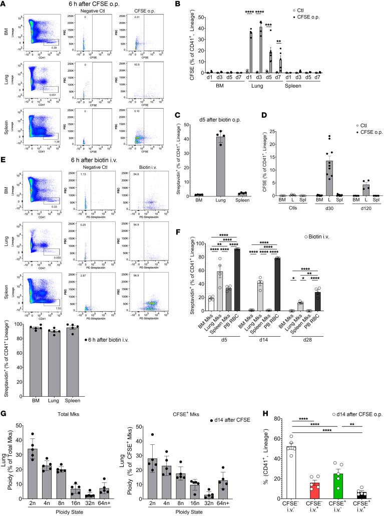
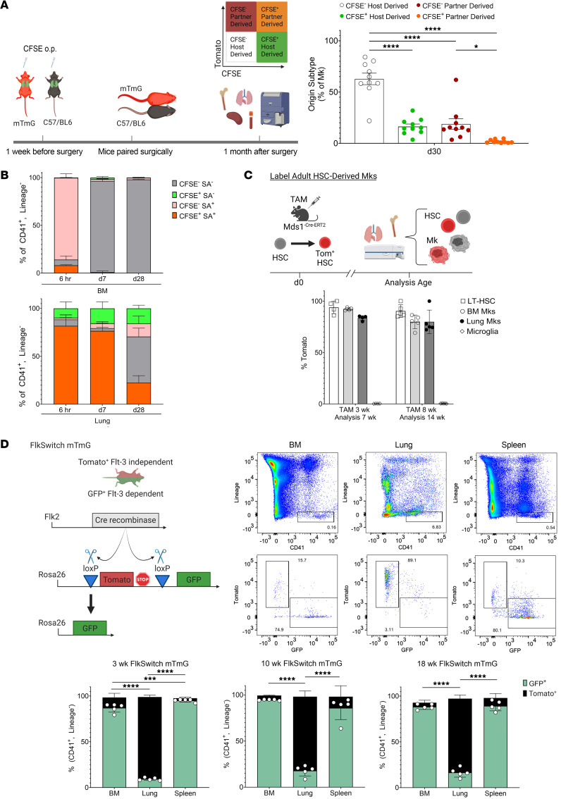
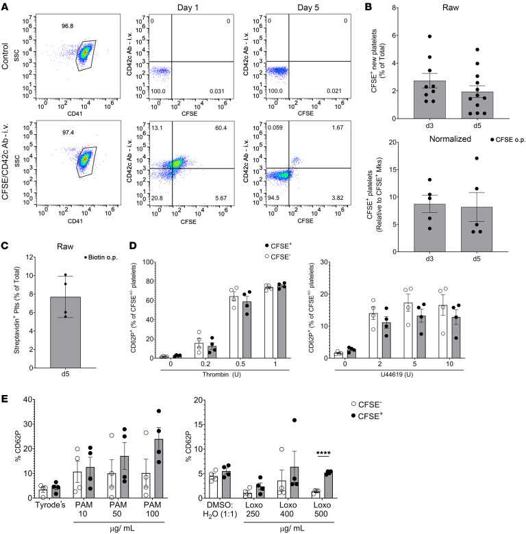
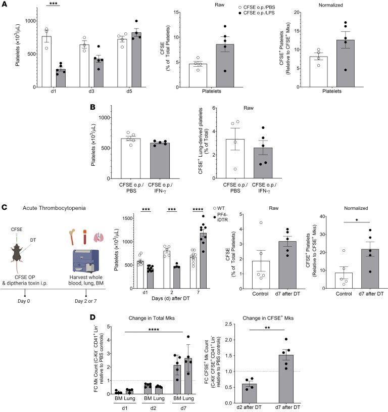
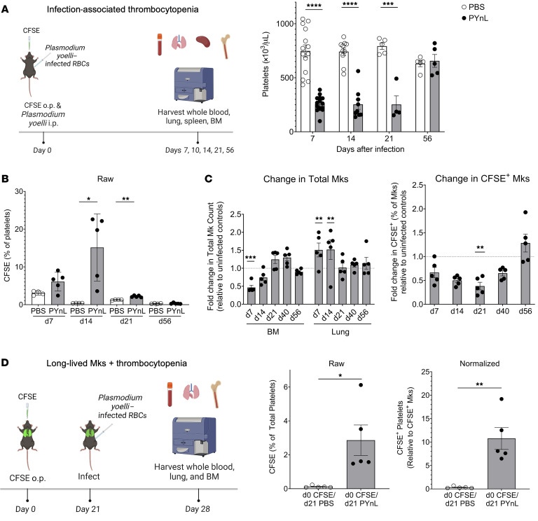
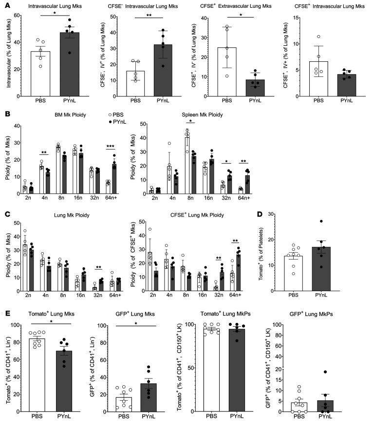
Lung megakaryocytes (Mks) are largely extravascular with an immune phenotype (1). Because bone marrow (BM) Mks are short lived, it has been assumed that extravascular lung Mks are constantly "seeded" from the BM. To investigate lung Mk origins and how origin affects their functions, we developed methods to specifically label lung Mks using CFSE dye and biotin delivered via the oropharyngeal route. Labeled lung Mks were present for up to 4 months, while BM Mks had a lifespan of less than 1 week. In a parabiosis model, lung Mks were partially replaced over 1 month from a circulating source. Unlike tissue-resident macrophages, using MDS1-Cre-ERT2 TdTomato mice, we found that lung Mks arose from hematopoietic stem cells. However, studies with FlkSwitch mTmG mice showed that lung Mks were derived from a Flt3-independent lineage that did not go through a multipotent progenitor. CFSE labeling to track lung Mk-derived platelets showed that approximately 10% of circulating platelets were derived from lung-resident Mks at steady state, but in sterile thrombocytopenia this was doubled (~20%). Lung-derived platelets were similarly increased in a malaria infection model (Plasmodium yoelii) typified by thrombocytopenia. These studies indicate that lung Mks arise from a Flt3- BM source, are long-lived, and contribute more platelets during thrombocytopenia.

Keywords: Hematology; Platelets.


Conflict of interest statement

Conflict of interest: The authors have declared that no conflict of interest exists.

Figures

Figure 1
Figure 1. Lung Mks are long-lived cells.
(A and B) CFSE or (C) biotin was delivered o.p. to mice and showed specific labeling of lung Mks, but not BM or splenic Mks. (A) CFSE 6 hours later (n = 4, 2 independent experiments), (B) CFSE on day 1 (d1) to day 7 (d7) (n = 4–5 per group from 2 independent experiments) and (C) on day 5 after biotin o.p. labeling (n = 4; results are representative of 2 independent experiments). (D) CFSE-labeled lung Mks were present up to 120 days after CFSE administration (n = 10 from 2 independent experiments for day 30; n = 4 for day 120 from 1 independent experiment). L, lung; Spl, spleen. (E) Representative flow cytometry and quantitation results 6 hours after biotin i.v. labeling of BM, splenic, and lung Mks, similar to A (n = 4; results are representative of 2 independent experiments). (F) Biotin-labeled lung but not BM or splenic Mks were present up to 28 days following i.v. delivery (n = 4; results are representative of 2 independent experiments). RBCs were used as a positive control. PB, peripheral blood. (G) Total lung Mks and CFSE+ Mks had similar ploidy (day 14 after CFSE) (n = 5, representative results are from 2 independent experiments). (H) CFSE and CFSE+ lung Mks had a similar intravascular (CD42d+) and extravascular (CD42d) distribution (CD42d given i.v.) (n = 5; results are representative of 2 independent experiments). Data indicate the mean ± SEM. *P < 0.05, **P < 0.01, ***P < 0.001, and ****P < 0.0001, by (B) multiple t tests with Holm-Šidák multiple-comparison correction, (D and F) 2-way ANOVA with Tukey’s multiple-comparison correction, and (H) 1-way ANOVA with Šidák multiple-comparison correction. Ctl, control; FSC, forward scatter.
Figure 2
Figure 2. Lung Mks are derived from BM HSCs.
(A) mTmG and Bl6/J mice were given CFSE o.p., and parabiosis surgeries performed 1 week later. Lung Mks were partially replaced by a BM source (n = 8–10 from 2 independent experiments). (B) Mice were given CFSE o.p. and biotin i.p., and 6 hours (n = 2 from 1 experiment), 7 days (n = 3 from 1 experiment), and 28 days (n = 5 from 1 experiment) later, BM and lung Mks were assessed. BM Mks were replaced within 7 days, but lung-resident Mks were detected at each time point. (C) MDS1-TdTomatoCre-ERT2 mice were treated with tamoxifen (TAM) to label HSCs and HSC-derived cells (n = 4–5; results are representative of 2 independent experiments). Lung Mks were HSC derived. (D) BM, splenic, and lung Mks in FlkSwitch mice were assessed at multiple ages (n = 5 per age time point; results are representative of 2 independent experiments) to determine whether Mk differentiation was Flt3 dependent. Lung Mks were largely Tomato+Flt3, indicating an HSC-to-Mk differentiation that was distinct from BM and splenic Flt3-dependent differentiation. Data indicate the mean ± SEM. *P < 0.05, ***P < 0.001, and ****P < 0.0001, by (B) 1-way ANOVA with Šidák multiple-comparison correction and (C and D) 2-way ANOVA with Tukey’s multiple-comparison correction and (A) 1-way ANOVA with Šidák multiple comparisons correction.
Figure 3
Figure 3. Lung-resident Mks produce platelets.
(A) Mice were simultaneously given CFSE o.p. and anti-CD42c platelet-labeling antibody i.p. Lung-derived platelets were quantified as CFSE+CD42c platelets by flow cytometry in representative plots. SSC, side scatter. (B) Results showed that 2%–5% of total platelets were lung derived (n = 9–12 from 3 independent experiments), and when normalized to CFSE+ Mks (n = 5; results are representative of 2 independent experiments), a maximum of approximately 10% of the platelets were lung-resident, Mk-derived. (C) CFSE data were validated by biotin delivered o.p. and by quantification of streptavidin-binding platelets (Plts) (n = 4; results are representative of 2 independent experiments). (D) CFSE+ lung-derived and CFSE platelets were agonist stimulated, and platelet activation was quantified by flow cytometry for CD62P surface expression. Lung-derived platelets and BM-derived platelets responded similarly to thrombin and U46619 (n = 4 per group; results are representative of 2 independent experiments). (E) Isolated platelets from mice treated with CFSE o.p. were stimulated with PAM3CSK4 (PAM) (10, 50, or 100 μg/mL) or Tyrode’s or with loxoribine (Loxo) (250, 400, or 500 μg/mL) or 1:1 DMSO/H2O, and CD62P surface expression was compared in either CFSE+ or CFSE platelets (n = 4 per group; results are representative of 2 independent experiments). Data indicate the mean ± SEM. *P < 0.05, **P < 0.01, ***P < 0.001, and ****P < 0.0001, by (D and E) multiple t tests with Holm-Šidák multiple-comparison correction.
Figure 4
Figure 4. Lung-resident Mks respond to acute thrombocytopenia.
(A) Mice were given CFSE o.p. and LPS or PBS i.p. LPS reduced platelet, but not Mk, counts and increased lung-derived platelets (n = 3–5 per group; results are representative of 2 independent experiments). (B) IFN-γ had no effect on platelet counts or CFSE+ platelets (n = 5 per group, representative shown from 2 independent experiments). (C) PF4Cre-iDTR mice treated with DT had reduced platelet counts (n = 5–9 per group from 2 independent experiments) and increased CFSE+ lung-derived platelets (n = 5 per group; results are representative of 2 independent experiments). DT treatment reduced BM and lung total Mks as well as CFSE+ Mks on day 2 after DT administration (n = 5 per group; results are representative of 2 independent experiments). (D) CFSE+ Mks relative to total Mks in control mice increased on day 7 after DT when Mk and platelet counts were recovered (n = 5 per group; results are representative of 2 independent experiments). Data indicate the mean ± SEM. *P < 0.05, **P < 0.01, ***P < 0.001, and ****P < 0.0001; (A and C) platelet count: multiple t tests with Holm-Šidák multiple-comparison correction; CFSE percentage and CFSE normalized: unpaired, 2-tailed t test; Mks: 2-way ANOVA with Tukey’s multiple-comparison correction; CFSE+ Mks: unpaired, 2-tailed t test; (B) unpaired, 2-tailed t tests; (D) Mks: 2-way ANOVA CFSE+ Mks: unpaired, 2-tailed t test.
Figure 5
Figure 5. Lung-resident Mks respond to infection-associated thrombocytopenia.
(A) Lung-resident, Mk-derived platelet counts were increased with PYnL infection–associated thrombocytopenia. Mice infected with PYnL had reduced platelet counts (n = 5–15 per group from 2 independent experiments) and (B) increased percentages of lung-derived CFSE+ platelets (n = 4–5 per group; results are representative of 2 independent experiments) in the first 2 weeks after infection. (C) Following PYnL infection, the number of BM Mks declined over the first 2 weeks, but the number of lung Mks increased (n = 4–5 per group per time point; results are representative of 2 independent experiments). The proportion of platelets that were CFSE+ was reduced early after infection, indicating an influx of new Mks from an extrapulmonary source. After infection recovery, the relative percentage of CFSE+ Mks in the lung were similar to that of control mice at the same time after CFSE. (D) Lung-resident Mks make platelets upon increased demand. Mice were treated with CFSE o.p. and infected 3 weeks later with PYnL. CFSE+ platelets were increased 1 week after infection (4 weeks after CFSE) (n = 5 per group; results are representative of 2 independent experiments). Data indicate the mean ± SEM. *P < 0.05, **P < 0.01, ***P < 0.001, and ****P < 0.0001; (A) platelets and (B) CFSE percentage of platelets and (C) CFSE+ Mks: multiple t tests with Holm-Šidák multiple-comparison correction; (C) total Mks: 2-way ANOVA with Holm-Šidák multiple-comparison correction; (D) CFSE percentage and normalized CFSE: unpaired, 2-tailed t test.
Figure 6
Figure 6. Mks migrate to the lung to respond to increased platelet demand.
(A) Mice were given CFSE o.p. and infected with PYnL, and on day 14, total Mks and CFSE+ and CFSE intravascular (CD42d i.v. -positive) and extravascular (CD42d i.v. –negative) Mks were quantified. The increase in total intravascular Mks was driven by CFSE extrapulmonary Mks. On day 14, there was a decrease in CFSE+ extra- and intravascular Mks (n = 5 per group, representative shown from 2 independent experiments). (B and C) With PYnL infection, there was an increase in higher ploidy CFSE+ Mks in (B) BM and spleen as well as in (C) lung, including CFSE+ Mks (n = 5 per group, representative shown from 2 independent experiments). (D) FlkSwitch mice infected with PYnL had no significant change in Tomato+Flt3 platelets on day 14 after infection, but (E) the percentage of lung Mks that were Tomato+Flt3 slightly declined and that of GFP+Flt3+ Mks increased slightly compared with uninfected controls. There was no change in MkPs, indicating an influx of mature Mks from the BM that was largely Tomato+Flt3 (n = 6–8 per group from 2 independent experiments). Data indicate the mean ± SEM. *P < 0.05, **P < 0.01, and ***P < 0.001, by unpaired, 2-tailed t test (A, D, and E) and multiple t tests with Holm-Šidák correction (B and C).

Comment in

  • The lung is a megakaryocyte outpost that can defend against thrombocytopenic attack

References

    1. Morrell CN, et al. The platelet napoleon complex-small cells, but big immune regulatory functions. Annu Rev Immunol. 2019;37:125–144. doi: 10.1146/annurev-immunol-042718-041607. - DOI - PubMed
    1. Morrell CN, et al. Emerging roles for platelets as immune and inflammatory cells. Blood. 2014;123(18):2759–2767. doi: 10.1182/blood-2013-11-462432. - DOI - PMC - PubMed
    1. Cloutier N, et al. Platelets can enhance vascular permeability. Blood. 2012;120(6):1334–1343. doi: 10.1182/blood-2012-02-413047. - DOI - PubMed
    1. Hilt ZT, et al. Platelet-derived β2M regulates monocyte inflammatory responses. JCI Insight. 2019;4(5):e122943. doi: 10.1172/jci.insight.122943. - DOI - PMC - PubMed
    1. Li C, et al. Thrombocytopenia independently leads to changes in monocyte immune function. Circ Res. 2024;134(8):970–986. doi: 10.1161/CIRCRESAHA.123.323662. - DOI - PMC - PubMed