. 2024 Sep 20;10(38):eadp2221.
doi: 10.1126/sciadv.adp2221. Epub 2024 Sep 20.

Molecular basis of TMED9 oligomerization and entrapment of misfolded protein cargo in the early secretory pathway

Affiliations

Molecular basis of TMED9 oligomerization and entrapment of misfolded protein cargo in the early secretory pathway

Le Xiao et al. Sci Adv. .

Abstract

Intracellular accumulation of misfolded proteins causes serious human proteinopathies. The transmembrane emp24 domain 9 (TMED9) cargo receptor promotes a general mechanism of cytotoxicity by entrapping misfolded protein cargos in the early secretory pathway. However, the molecular basis for this TMED9-mediated cargo retention remains elusive. Here, we report cryo-electron microscopy structures of TMED9, which reveal its unexpected self-oligomerization into octamers, dodecamers, and, by extension, even higher-order oligomers. The TMED9 oligomerization is driven by an intrinsic symmetry mismatch between the trimeric coiled coil domain and the tetrameric transmembrane domain. Using frameshifted Mucin 1 as an example of aggregated disease-related protein cargo, we implicate a mode of direct interaction with the TMED9 luminal Golgi-dynamics domain. The structures suggest and we confirm that TMED9 oligomerization favors the recruitment of coat protein I (COPI), but not COPII coatomers, facilitating retrograde transport and explaining the observed cargo entrapment. Our work thus reveals a molecular basis for TMED9-mediated misfolded protein retention in the early secretory pathway.


Figures

Fig. 1.
Fig. 1.. Structures of TMED9 oligomers.
(A) Domain organization of TMED9. TM, transmembrane helix; CT, cytoplasmic tail. Residue numbers are shown. (B) Cryo-EM map of TMED9 octamer with TMED chains in cyan, green, and magenta, and lipid density in gray. For the magenta-colored chains, the coiled coil region is disordered. (C) A representative TMED9 subunit structure labeled with the color scheme in (A). (D) TMED9 octamer model shown with the color scheme in (B). (E) The tetrameric substructures of TMED9 TM domain (top) and the trimeric substructures of TMED9 coiled coli domain (bottom). In the schematic diagrams at right, chain identifications are labeled. The eye-shaped and triangular symbols are C2 and C3 axes, respectively. (F to H) TMED9 dodecamer model in two orientations [(F) and (G)] and its schematic (H). The four extra TMED9 chains in comparison with the octamer are shown in magenta and pink. Each color in the schematic of the TM arrangement represents a trimer in the coiled coil domain. (I) The cryo-EM density and the location of the fitted PIP lipid (indicated by stick figures in the schematic).
Fig. 2.
Fig. 2.. Interactions within TMED9 oligomers.
(A to C) Ile/Leu-mediated hydrophobic interactions (in yellow) within the TM tetramer and coiled coil trimer of TMED9. PIP is in magenta. (D) Hydrophilic interactions within the TMED9 coiled coil trimer. (E) Hydrophilic interactions between TMED9 coiled coil trimers. (F) Hydrophilic interactions between PIP and TMED9 TM domain, as protein surface charge overview (left, in plane of bilayer) and as helical ribbons (right) viewed looking toward the monomeric N termini. (G) Gel filtration profile of WT and mutant TMED9. The vertical dashed line marks the peak elution of WT TMED9. (H) Mass photometry profile of WT and mutant TMED9. The vertical dashed line marks the expected position of octamers of WT TMED9. Residues mutated here are within red boxes in (D) to (F).
Fig. 3.
Fig. 3.. Mapping of the interaction between TMED9 and MUC1-fs.
(A) Schematic of MUC1 and MUC1-fs constructs. SP, signal peptide; R1–Rn, repeats in MUC1; F1–Fn, repeats in MUC1-fs; OR, ordered region; TM, transmembrane domain; CD, cytoplasmic domain; MBP, maltose-binding protein; Flag, FLAG tag. (B) FLAG co-IP of FLAG-tagged MUC1-fs WT and truncation constructs and coexpressed TMED9-Myc. MUC1-fs constructs ran as monomers and/or dimers on reducing SDS-PAGE and immunoblots. Lys, lysate; FT, flow through; IP, IP’ed beads. Bands corresponding to the MW of the MUC1-fs constructs are in red boxes for the IP lane. Some MUC1-fs constructs ran partly as a dimer even in reducing SDS-PAGE, leading to the additional bands. (C) FLAG co-IP of FLAG-tagged MBP (negative control, left) or FLAG-tagged MBP-OR (right) with coexpressed WT and single-domain deletions of Myc-tagged TMED9.
Fig. 4.
Fig. 4.. Mode of interaction between MUC1-fs OR and TMED9 GOLD domain.
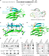
(A) Sequence and predicted secondary structure of the OR of MUC1-fs. (B) AlphaFold–predicted top-ranked OR-GOLD domain complex model (left) and overlay of the five predicted models (right). In the top-ranked model, TMED9 GOLD domain is in green and MUC1-fs OR is in cyan, and the predicted disulfide bonds are in yellow. (C) Interactions are shown between the β1 strand of OR (cyan for carbon atoms) and the β2 strand of GOLD (green for carbon atoms). Other atoms are shown in blue for nitrogen, red for oxygen, and yellow for sulfur. Main chain hydrogen bonds are indicated by black dotted lines. (D) FLAG co-IP of FLAG-tagged MBP-OR and Myc-tagged TMED9 WT and mutants. Lys, lysate; FT, flow through; IP, IP’ed. beads. (E) OR-GOLD domain complex model with C925 of OR mutated to Ser, C49, and C121 of the GOLD domain mutated to Ser, and E53 of the GOLD domain mutated to Cys, showing the proximity between C911 of OR and C53 of the GOLD domain for possible disulfide bond formation. (F) FLAG co-IP of FLAG-tagged C911-only MBP-OR and E53C-only Myc-tagged TMED9. Lys, lysate; FT, flow through; IP, IP’ed. beads. The red dashed boxes indicate disulfide-linked TMED9 and MBP-OR under nonreducing conditions (−DTT) absent under reducing conditions (+DTT). One and two open black circles, respectively, mark the MBP-OR monomer and dimer bands. One and two open black triangles respectively mark TMED9 monomer and dimer bands. MBP-OR migrates as both monomer and dimer even under reducing conditions, for unknown reasons.
Fig. 5.
Fig. 5.. Interaction of TMED9 with COPI but not COPII components.
(A) Volcano plot of the TMED9 interactome (Myc-TMED9 + MUC1-fs) shows preferential binding of TMED9 (green dot) to COPI proteins (red dots), but not to COPII proteins (purple dots), which did not pass the significance threshold for enrichment compared to the negative control (empty vector + MUC1-fs). (B) Myc-tagged TMED9 co-IP’ed. COPB2 (COPI protein) but not SEC13 (COPII protein). β-Actin is shown as a loading control in the lysate and as a negative control in the pulldown. Blots are representative of three independent experiments. (C) Myc-tagged TMED9 mutant R223E coimmunoprecipitated less endogenous COPB2 than WT TMED9. (D) GFP-tagged SEC23a coimmunoprecipitated mutant and WT TMED9. **P < 0.05. ns., not significant.
Fig. 6.
Fig. 6.. Schematic of the function of TMED9 in mutant cargo accumulation in COPI compartments.
Newly expressed MUC1-fs interacts with the GOLD domain of TMED9 (step 1) in the ER and is incorporated into the COPII compartment (vesicles or tubules) (step 2), which mediate anterograde transport and release into the cis-Golgi (step 3). In the cis-Golgi, misfolded MUC1-fs can further oligomerize because of the more oxidizing environment, which may in turn increase interaction of the TMED9–MUC1-fs complex with COPI coatomers (step 4). The tripartite COPI-TMED9–MUC1-fs complexes may then attempt to undergo retrograde transport back toward the ER (step 5). The higher-order oligomerization state of the TMED9–MUC1-fs complex may inhibit its reentry into the ER, or it may prevent interactions between TMED9 and COPII coatomers (step 6). As a result, the misfolded, aggregated MUC1-fs is trapped in COPI-predominant compartments in complex with higher-order oligomeric TMED9.

References

    1. Higuchi-Sanabria R., Frankino P. A., Paul J. W. III, Tronnes S. U., Dillin A., A futile battle? Protein quality control and the stress of aging. Dev. Cell 44, 139–163 (2018). - PMC - PubMed
    1. Dvela-Levitt M., Shaw J. L., Greka A., A rare kidney disease to cure them all? Towards mechanism-based therapies for proteinopathies. Trends Mol. Med. 27, 394–409 (2021). - PubMed
    1. Dvela-Levitt M., Kost-Alimova M., Emani M., Kohnert E., Thompson R., Sidhom E.-H., Rivadeneira A., Sahakian N., Roignot J., Papagregoriou G., Small molecule targets TMED9 and promotes lysosomal degradation to reverse proteinopathy. Cell 178, 521–535.e23 (2019). - PubMed
    1. Thomas G., Horwich A., Chemical strike against a dominant-inherited MUC1-frameshifted protein associated with progressive kidney disease. Trends Mol. Med. 25, 821–823 (2019). - PMC - PubMed
    1. Dhar P., McAuley J., The role of the cell surface mucin MUC1 as a barrier to infection and regulator of inflammation. Front. Cell. Infect. Microbiol. 9, 117 (2019). - PMC - PubMed

LinkOut - more resources