. 2024 Sep 20;10(38):eadn7724.
doi: 10.1126/sciadv.adn7724. Epub 2024 Sep 20.

Generation and characterization of giant panda induced pluripotent stem cells

Affiliations

Generation and characterization of giant panda induced pluripotent stem cells

Yuliang Liu et al. Sci Adv. .

Abstract

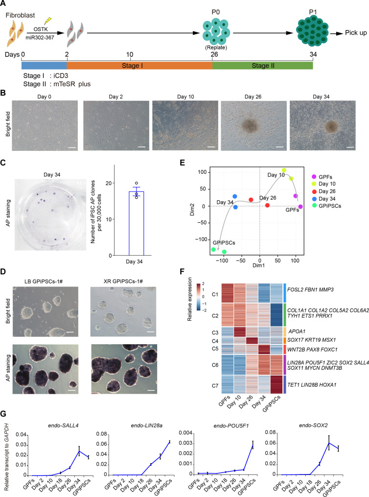
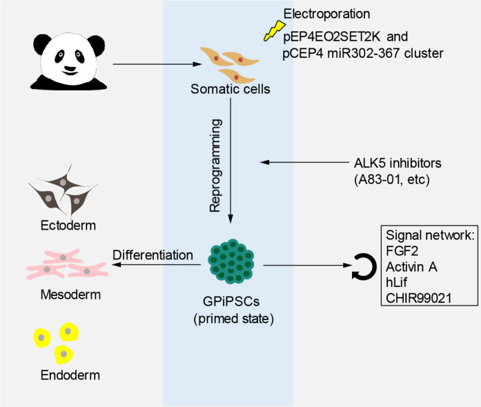
The giant panda (Ailuropoda melanoleuca) stands as a flagship and umbrella species, symbolizing global biodiversity. While traditional assisted reproductive technology faces constraints in safeguarding the genetic diversity of giant pandas, induced pluripotent stem cells (iPSCs) known for their capacity to differentiate into diverse cells types, including germ cells, present a transformative potential for conservation of endangered animals. In this study, primary fibroblast cells were isolated from the giant panda, and giant panda iPSCs (GPiPSCs) were generated using a non-integrating episomal vector reprogramming method. Characterization of these GPiPSCs revealed their state of primed pluripotency and demonstrated their potential for differentiation. Furthermore, we innovatively formulated a species-specific chemically defined FACL medium and unraveled the intricate signaling pathway networks responsible for maintaining the pluripotency and fostering cell proliferation of GPiPSCs. This study provides key insights into rare species iPSCs, offering materials for panda characteristics research and laying the groundwork for in vitro giant panda gamete generation, potentially aiding endangered species conservation.


Figures

Fig. 1.
Fig. 1.. Derivation of giant panda pluripotent stem cell.
(A) Schematic of reprogramming from fibroblasts to GPiPSCs. (B) Morphology of transduced GPFs on day 0, day 2, day 10, day 26, and day 34. Scale bars, 200 μm. (C) Left: AP staining of iPSCs generated from GPF cells in 12-well plates in mTeSR Plus medium. Right: Reprogramming efficiencies of AP-positive cells. n = 3. (D) Morphology and AP of XR (Xingrong, XX) GPiPSCs-1#and LB (Loubao, XY) GPiPSCs-1# in mTeSR Plus medium. Scale bars, 100 μm. (E) t-SNE analysis of the transcriptomes of GPiPSC induction. (F) Heatmaps of the transcriptomes during GPiPSC induction. (G) qRT-PCR validation of pluripotency genes expression during GPiPSC induction. For (C) and (G), the quantitative data represent the mean ± SE; n = 3 independent experiments.
Fig. 2.
Fig. 2.. Enhancing giant panda pluripotent stem cell induction efficiency with ALK5 inhibitors.
(A) Schematic of reprogramming from fibroblasts to GPiPSCs with different compounds. (B) Table of tested compounds and their known targets. (C) AP staining of GPiPSCs reprogrammed by different compounds in 12-well plates in mTeSR Plus medium. (D) Reprogramming efficiencies of different compounds. n = 3. (E) Comparison of transcriptomes of cells reprogrammed by iCD3 and iCD3 + A83-01 in day 18, with differentially expressed genes (DEGs) [>2 times, false discovery rate (FDR) < 0.001, log2TPM > 1]. (F) Morphology and AP staining of XR (Xingrong, XX) GPiPSCs-4#, 5# reprogrammed by iCD3 + A83-0 in mTeSR Plus medium. Scale bars, 100 μm. For (D), the quantitative data represent the mean ± SE; n = 3 independent experiments. ***P < 0.001.
Fig. 3.
Fig. 3.. Characterization of pluripotent stem cells in giant panda.
(A) Immunostaining of pluripotency marker (SALL4, OCT4, and LIN28A) in GPiPSCs in mTeSR Plus medium. Scale bars, 100 μm. (B) Growth curve of XR GPiPSCs-1# and LB GPiPSCs-1# at passages 10 and 30 in mTeSR Plus medium. (C) Karyotype analysis of XR GPiPSCs-1# and LB GPiPSCs-1# at passage 30 in mTeSR Plus medium. Normal: 42, XX with 20 matched pairs of autosomes and X/X paired sex chromosomes; XY with 20 pairs of matched autosomes and X/Y unpaired sex chromosomes. (D) Scatterplots of transcriptomes of GPiPSCs and GPFs, with DEGs (>2 times, FDR < 0.001, log2TPM > 1). (E) Principal components analysis of GPiPSCs in comparison to iPSCs derived from other species. Each dot represents one dataset. (F) Heatmaps of the transcriptomes between GPiPSCs and other species. (G) Gene Ontology (Biological Process) [GO(BP)] analyses of significantly up-regulated gene (cluster 10) in GPiPSCs. DAPI, 4A,6-diamidino-2-phenylindole; ncRNA, noncoding RNA; tRNA, transfer RNA.
Fig. 4.
Fig. 4.. The primed state of giant panda pluripotent stem cells.
(A) The Venn diagrams identify genes co–up-regulated in the naïve or primed state in human and mouse PSCs, with DEGs (>2 times, FDR < 0.001, log2TPM > 1). (B) The heatmap shows correlation coefficients of gene expression representing the pluripotent state. (C) qRT-PCR validation of pluripotent marker genes (POU5F1, SOX2, and SALL4), primed state marker genes (ZIC2, SOX11, and DNMT3B), and naïve state marker genes (KLF4, TEF3, and DNMT3L) in LB GPiPSCs-1# (LB-1#), LB GPiPSCs-2# (LB-2#), XR GPiPSCs-1# (XR-1#), and XR GPiPSCs-2# (XR-2#). (D) Bisulfite sequencing analysis of the DNA CpG methylation statuses of the primed state marker gene ZIC2 and the naïve state marker gene TEF3 promoter loci in GPiPSCs and GPFs. (E) Immunostaining for hypermethylation of H3 lysine 27me3 (H3K27me3) in mTeSR Plus medium. Scale bars, 10 μm. For (C), the quantitative data represent means ± SE; n = 3 independent experiments. *P < 0.05, **P < 0.01, and ***P < 0.001.
Fig. 5.
Fig. 5.. Potentiation of differentiation in giant panda pluripotent stem cells.
(A) EBs derived from GPiPSCs in differentiation medium. Images were taken at days 4, 8, 12, 16, and 20. Scale bars, 200 μm. (B) qRT-PCR validation of selected genes at days 4, 8, 12, 16, and 20 of EB differentiation. (C) Immunostaining for markers of endoderm (SOX17), mesoderm (SMA and GATA4), and ectoderm (β3-TUBULIN). Scale bars, 50 μm. (D) Images of teratoma derived from GPiPSCs at 45 days and microscopic images of H&E-stained sections of teratoma. Scale bars, 50 μm. For (B), the quantitative data represent means ± SE; n = 3 independent experiments. *P < 0.05, **P < 0.01, and ***P < 0.001.
Fig. 6.
Fig. 6.. Signaling pathways maintaining pluripotency in giant panda pluripotent stem cells.
(A) Morphology and fluorescence images of POU5F1-2A-EGFP GPiPSCs in mTeSR Plus medium and FACL medium. Scale bars, 100 μm. (B) Growth curve of GPiPSCs in mTeSR Plus medium and FACL medium. (C) Scatterplots comparing the global gene expression of GPiPSCs in mTeSR Plus medium and FACL medium, with DEGs (>4 times, FDR < 0.001, log2TPM > 1). (D) Morphology and fluorescence images of GPiPSCs culture in FACL medium and FACL medium minus different components. Scale bars, 100 μm. (E) Growth curve of giant panda PSCs in FACL medium and FACL medium minus different components. (F) Scatterplots comparing the global gene expression of giant panda PSCs in FACL medium and FACL medium minus CHIR99021, with DEGs (>2 times, FDR < 0.001, log2TPM > 1). (G) GO analyses of significantly up-regulated gene in FACL medium minus CHIR99021. (H) Heatmap of the transcriptome data of lineage marker genes between FACL medium and FACL medium minus CHIR99021. (I) Fluorescence images of pluripotent marker proteins (SALL4 and LIN28A) in FACL medium and FACL medium minus CHIR99021. Scale bars, 100 μm. (J) Heatmaps of the transcriptome data of CKIs between FACL medium and FACL medium minus CHIR99021, phycoerythrin (PE-A). (K) Result of cell cycle in FACL medium and FACL medium minus CHIR99021. For (B), (E), and (K), the quantitative data represent means ± SE; n = 3 independent experiments. **P < 0.01.
Fig. 7.
Fig. 7.. Proposed model for generation and characterization of iPSCs in giant pandas.
Primary fibroblast cells are isolated from the giant panda, and GPiPSCs are generated using a non-integrating episomal vector reprogramming method, while ALK5 inhibitors can enhance GPiPSC induction efficiency. GPiPSCs were in the primed state when cultured in FACL medium and have the ability to differentiate into the three germ layers.

References

    1. Wei F., Fan H., Hu Y., Ailuropoda melanoleuca (Giant Panda). Trends Genet. 36, 68–69 (2020). - PubMed
    1. Kang D., A review of the impacts of four identified major human disturbances on the habitat and habitat use of wild giant pandas from 2015 to 2020. Sci. Total Environ. 763, 142975 (2021). - PubMed
    1. Swaisgood R. R., Wang D., Wei F., Panda downlisted but not out of the woods. Conserv. Lett. 11, e12355 (2018).
    1. Kong L., Xu W., Xiao Y., Pimm S. L., Shi H., Ouyang Z., Spatial models of giant pandas under current and future conditions reveal extinction risks. Nat. Ecol. Evol. 5, 1309–1316 (2021). - PubMed
    1. Zhang M., Hou R., Zheng H., Zhu Q., Zhang Z., Liu Y., Establishment and cryopreservation of a giant panda skin fibroblast cell line. Chin. J. Biol. 40, 61–67 (2005).

Publication types