

Quantitative MRI Measures and Cognitive Function in People With Drug-Resistant Juvenile Myoclonic Epilepsy
- PMID: 39303180
- PMCID: PMC11446167
- DOI: 10.1212/WNL.0000000000209802
Quantitative MRI Measures and Cognitive Function in People With Drug-Resistant Juvenile Myoclonic Epilepsy
Abstract
Background and objectives: Neuroimaging studies have so far identified structural changes in individuals with juvenile myoclonic epilepsy (JME) when compared with controls. However, the underlying mechanisms of drug-resistant JME remain unknown. In this study, we aimed at characterizing the structural underpinnings of drug-resistant JME using MRI-derived cortical morphologic markers.
Methods: In this prospective cross-sectional 2-center study, T1-weighted MRI and neuropsychological measures of verbal memory and executive function were obtained in individuals with drug-resistant and drug-responsive JME recruited from epilepsy outpatient clinics and healthy controls. We performed vertexwise measurements of cortical thickness, surface area, and local gyrification index (LGI). Vertexwise group comparisons were corrected for multiple comparisons at a familywise error (FWE) of 0.05. The neuropsychological profile of disease subgroups was analyzed through principal component analysis.
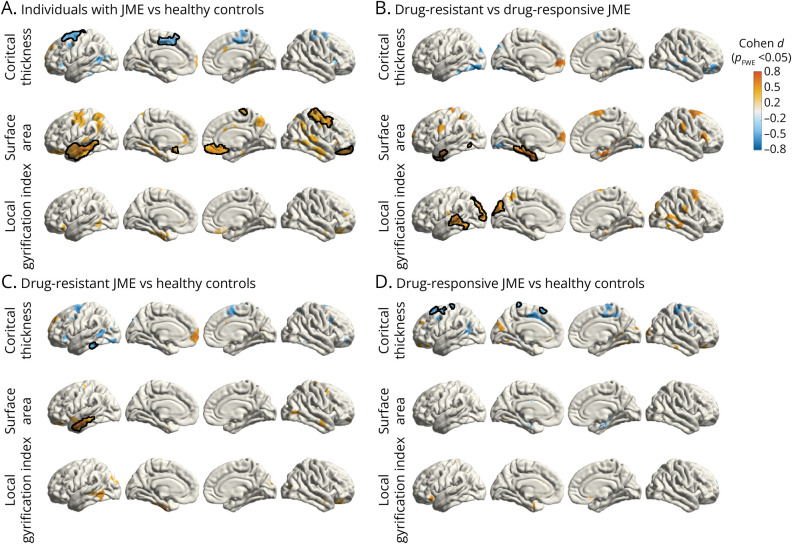
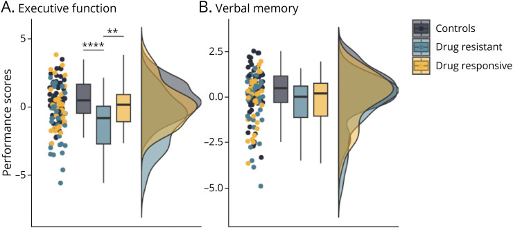
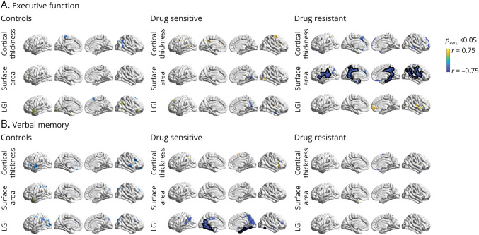
Results: We studied 42 individuals with drug-resistant JME (mean age 29 ± 11 years, 50% female), 37 with drug-responsive JME (mean age 34 ± 10, years, 59% female), and 71 healthy controls (mean age 21 ± 9 years, 61% female). Surface area was increased in participants with drug-resistant JME in the left temporal lobe (Cohen d = 0.82 [-0.52 to -1.12], pFWE < 0.05) when compared with the drug-responsive group. Although no cortical thickness changes were observed between disease subgroups, drug-resistant and drug-sensitive participants showed discrete cortical thinning against controls (Cohen d = -0.42 [-0.83 to -0.01], pFWE < 0.05; Cohen d = -0.57 [-1.03 to -0.11], pFWE < 0.05, respectively). LGI was increased in the left temporal and occipital lobes in drug-resistant participants (Cohen d = 0.60 [0.34-0.86], pFWE < 0.05) when contrasting against drug-sensitive participants, but not controls. The composite executive function score was reduced in drug-resistant individuals compared with controls and drug-sensitive individuals (-1.74 [-2.58 to -0.90], p < 0.001 and -1.29 [-2.25 to -0.33], p < 0.01, respectively). Significant correlations were observed between executive function impairment and increased surface area in the precuneus and medial prefrontal regions (r = -0.79, pFWE < 0.05) in participants with drug-resistant JME.
Discussion: We identified a developmental phenotype in individuals with drug-resistant JME characterized by changes in cortical surface area and folding complexity, the extent of which correlates with executive dysfunction. No association was observed between cortical thickness and disease severity. Our findings support a neurodevelopmental basis for drug resistance and cognitive impairment in JME.
Conflict of interest statement
B. Crespo Pimentel was supported by a scholarship from the Austrian Society of Epileptology, which was not directly related to this project. E. Trinka has received research funding from Austrian Science Fund grant KLI 969 and reports personal fees from EVER Pharma, Marinus, Argenx, Arvelle/Angelini, Medtronic, Bial-Portela & Ca, NewBridge, GL Pharma, GlaxoSmithKline, Hikma, Boehringer Ingelheim, LivaNova, Eisai, UCB, Biogen, Genzyme Sanofi, GW Pharmaceuticals/Jazz, and Actavis, outside the submitted work; his institution has received grants from Biogen, UCB Pharma, Eisai, Red Bull, Merck, Bayer, the European Union, FWF Osterreichischer Fond zur Wissenschaftsforderung, Bundesministerium für Wissenschaft und Forschung, and Jubilaumsfond der Österreichischen Nationalbank, outside the submitted work. M. Kronbichler has received honoraria from Biocodex, Bial, GE, GSK, LivaNova, Eisai, UCB, and Jazz Pharmaceuticals, and has received research funding from MRC, the Wellcome Trust (grant 079474), ER-UK, the Henry Smith Foundation (grant 20133416), and the Epilepsy Society. Britta Wandschneider has received research funding from the Henry Smith Foundation (grant 20133416) and has received salary support from the German Research Foundation (WA3135/1-1). The remaining authors have no conflicts of interest. Go to
Figures
References
-
- Riney K, Bogacz A, Somerville E, et al. . International League Against Epilepsy classification and definition of epilepsy syndromes with onset at a variable age: position statement by the ILAE Task Force on Nosology and Definitions. Epilepsia. 2022;63(6):1443-1474. doi:10.1111/epi.17240 - DOI - PubMed
Publication types
MeSH terms
LinkOut - more resources
Full Text Sources
Medical