. 2024 Oct;300(10):107784.
doi: 10.1016/j.jbc.2024.107784. Epub 2024 Sep 18.

Protein phosphatase PP2Cα S-glutathionylation regulates cell migration

Affiliations

Protein phosphatase PP2Cα S-glutathionylation regulates cell migration

Dhanushika S K Kukulage et al. J Biol Chem. 2024 Oct.

Abstract

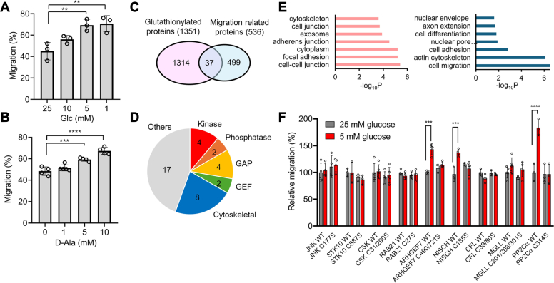
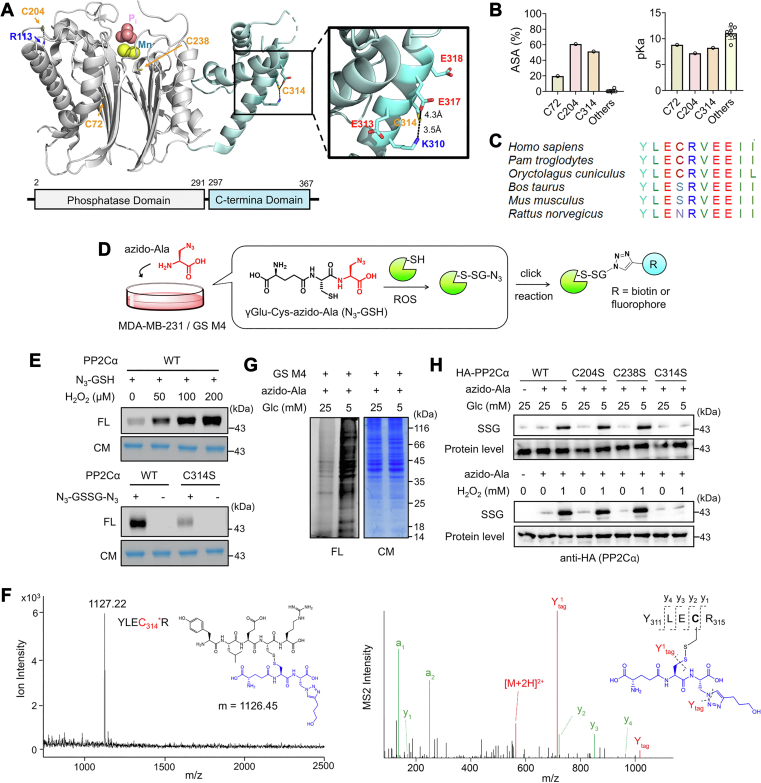
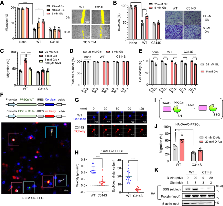
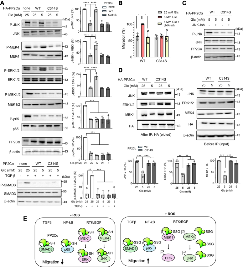
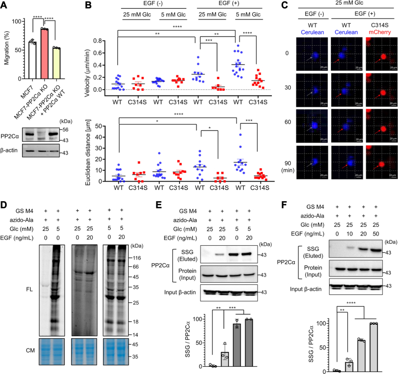
Redox signaling is a fundamental mechanism that controls all major biological processes partly via protein cysteine oxidations, including S-glutathionylation. Despite over 2000 cysteines identified to form S-glutathionylation in databases, the identification of redox cysteines functionally linked to a biological process of interest remains challenging. Here, we demonstrate a strategy combining glutathionylation proteomic database, bioinformatics, and biological screening, which resulted in the identification of S-glutathionylated proteins, including PP2Cα, as redox players of cell migration. We showed that PP2Cα, a prototypical magnesium-dependent serine/threonine phosphatase, is susceptible to S-glutathionylation selectively at nonconserved C314. PP2Cα glutathionylation causes increased migration and invasion of breast cancer cell lines in oxidative stress or upon hydrogen peroxide production. Mechanistically, PP2Cα glutathionylation modulates its protein-protein interactions, activating c-Jun N-terminal kinase and extracellular signal-regulated kinase pathways to elevate migration and invasion. In addition, PP2Cα glutathionylation occurs in response to epidermal growth factor, supporting a serine/threonine phosphatase PP2Cα as a new redox player in growth factor signal transduction.

Keywords: PP2Cα phosphatase; cell migration; epidermal growth factor; glutathionylation; reactive oxygen species; redox signaling.


Conflict of interest statement

Conflicts of interests The authors declare that they have no conflicts of interest with the contents of this article.

Figures

Figure 1
Figure 1
A strategy to identify functional cysteines regulating cell migration via S-glutathionylation. Proteomic data, bioinformatics, and biological screening (i.e., cell migration) were combined to narrow down regulatory cysteines for cell migration. A–B, low glucose or D-amino acid oxidase (DAAO)-induced H2O2 increases cell migration. MCF7 cells were incubated with decreasing concentrations of glucose (n = 3) (A) or MCF7 cells expressing DAAO were incubated with increasing concentrations of D-Ala for 24 h (n = 4) (B). C, Cytoscape bioinformatic analysis comparing glutathionylated proteins and migration-related proteins. D, functional categories of 37 proteins belonging to “S-glutathionylation” and “migration” groups, including GTPase-activating protein (GAP) and guanine nucleotide exchange factor (GEF). E, gene ontology (GO) analysis of the identified 37 proteins. F, cell migration screening of nine selected proteins. MDA-MB-231 cells expressing protein WT or cysteine-to-serine mutant (C/S) were subjected to high or low glucose conditions. The migration was measured using the wound-healing assay and normalized by the average migration levels of each protein WT at 25 mM glucose (n = 3). ∗STK10, Rab21, and MGLL with cysteine numbers are from mouse, and all others are from human. Data represent the mean ± SD. The statistical difference was analyzed by one-way ANOVA and Tukey’s post hoc test (A, B) or two-way ANOVA followed by Tukey’s post hoc test (F), where ∗p < 0.03, ∗∗p < 0.002, ∗∗∗p < 0.0002, ∗∗∗∗p < 0.0001.
Figure 2
Figure 2
PP2Cα is susceptible to glutathionylation at non-conserved Cys 314.A, PP2Cα structure. A model shows phosphate (Pi) and Mn2+ in the active site and locations of three relatively surface-exposed cysteines (C72, C204, and C314). B, comparisons of pKa and accessible surface area (ASA) for cysteines in PP2Cα. C, the amino acid sequence alignment around C314 among different species, showing limited conservation of C314. D, a scheme of a clickable glutathione approach for analyzing S-glutathionylation. Cells expressing GS M4 are incubated with azido-Ala, which enables biosynthesis of clickable glutathione (azido-glutathione, N3-GSH). N3-GSH forms S-glutathionylation in oxidative stress. Alternatively, purified protein can form glutathionylation in the presence of N3-GSH, which is analyzed after the click reaction. E, glutathionylation of purified PP2Cα in vitro. Purified PP2Cα was incubated with N3-GSH and H2O2 for 15 min (top) or oxidized azido-glutathione (N3-GSSG-N3) for 30 min (bottom). PP2Cα glutathionylated by azido-glutathione was conjugated with rhodamine-alkyne via click chemistry and visualized by Coomassie stain (CM, protein level) and fluorescence (FL, SSG level) (n = 3). F, MALDI-TOF and MS/MS analyses of glutathionylated peptides in PP2Cα. Purified PP2Cα glutathionylated by azido-glutathione was click-conjugated by biotin-DADPS-alkyne, enriched by streptavidin-agarose, and digested by trypsin on beads. Glutathionylated peptides were eluted and analyzed by MALDI-TOF (left) and LC-MS/MS (right), finding a glutathionylated peptide at C314 (YLEC314∗R, m/z 1127). In the MS2 spectrum, all y ions were found with additional ions (Y1tag and Ytag) resulting from fragmentation in glutathione modification (n = 3). G, global glutathionylation in MDA-MB-231. MDA-MB-231 cells expressing GS M4 (MDA-MB-231/GS M4) were stimulated in low or high glucose conditions. After the click reaction of lysates with rhodamine-alkyne, proteins were analyzed by fluorescence and Coomassie stain (n = 3). H, PP2Cα glutathionylation in MDA-MB-231. MDA-MB-231/GS M4 cells expressing HA-PP2Cα WT or C/S mutant were incubated in low or high glucose conditions for 24 h or with H2O2 for 15 min. After the click reaction of lysates with biotin-alkyne, glutathionylated PP2Cα was probed by Western blot before (protein level) and after (SSG) enrichment by streptavidin-agarose (n = 3). Data are representative of three independent experiments.
Figure 3
Figure 3
PP2Cα C314 glutathionylation increases cell migration and invasion. A–E, analyses of MDA-MB-231 cells expressing PP2Cα WT or C314S for migration, invasion, and viability. MDA-MB-231 cells expressing PP2Cα WT or C314S were incubated in different glucose concentrations. A, the wound-healing migration assay for 36 h (n = 3). The yellow color indicates the area without cells. The scale bar represents 0.5 mm. B, transwell-invasion assay for 24 h (n = 3). The scale bar represents 0.1 mm. C, the wound-healing migration assay upon adding N-acetylcysteine (NAC) for 24 h (n = 3). D, total cell numbers after 24 h (n = 3). E, cell viability after 24 h (n = 3). FH, tracking migration of individual cells. MDA-MB-231 cells expressing PP2Cα WT/Cerulean (blue) or PP2Cα C314S/mCherry (red) were combined and incubated in low glucose (5 mM) with EGF for 2 h. Cells were monitored by fluorescence every 10 min (0–120 min). The images were combined to analyze the migration of individual cells (n = 10–12). F, bicistronic plasmid maps (top) and the confocal image with migration tracks of individual cells (bottom). Cells visible during the entire time frame were analyzed. Red boxes show red cells, whereas blue boxes show blue cells. In each box, migration tracks over 2 h are shown by tails. The scale bar represents 40 μm. G, representative images of individual cells at different time points, showing the migration. The scale bar represents 20 μm. H, migration velocity and distance. Individual cells were analyzed and shown in a dot plot. I, the PROX-D system. DAAO is fused to the protein of interest (POI) (i.e., PP2Cα), and D-Ala incubation produces H2O2 in proximity to POI, which causes POI oxidations. JandK, cell migration and PP2Cα glutathionylation induced by PROX-D. DAAO-PP2Cα WT or C314S was expressed in MDA-MB-231 cells. Cell migration (n = 3) (J) and PP2Cα glutathionylation (n = 3) (K) were measured after adding D-Ala to cells in 3 mM glucose for 24 h. Data represent the mean ± SD. The statistical difference was analyzed by two-way ANOVA and Tukey’s post hoc test (A–E, and J) or two-tailed Student’s unpaired t test (H), where ∗p < 0.03, ∗∗p < 0.002, ∗∗∗p < 0.0002, ∗∗∗∗p < 0.0001.
Figure 4
Figure 4
PP2Cα C314 glutathionylation activates JNK and ERK pathways by selectively altering protein–protein interactions.A, activation of JNK, ERK1/2, and MEK4 upon PP2Cα glutathionylation. MDA-MB-231 cells expressing PP2Cα WT or C314S were incubated in low or high glucose for 6 h, and phosphorylation levels of PP2Cα substrates were analyzed by western blots (n = 3). BC, JNK activation is responsible for increased cell migration. The wound-healing migration for 24 h (n = 3) (B) and JNK phosphorylation after 6 h (n = 3) (C) in cells expressing PP2Cα WT or C314S upon the addition of JNK-inhibitor (JNK-Inh). D, PP2Cα glutathionylation induces its dissociation from JNK, ERK1/2, and MEK4. After incubation of cells in low or high glucose for 6 h, co-immunoprecipitation was used to monitor their binding interactions by western blots (n = 3). E, a model for cell migration induced by PP2Cα glutathionylation. In a nonstressed condition, PP2Cα binds to its substrates for their dephosphorylation, which suppresses cell migration. However, H2O2 or ROS induced by low glucose causes PP2Cα glutathionylation at C314, which dissociates PP2Cα from selected signaling substrates, such as JNK, ERK1/2, and MEK4. The dissociation increases phosphorylation levels of JNK, ERK1/2, and MEK4, which causes increased cell migration via the JNK-paxillin pathway. Data show the mean ± SD or representative of three independent experiments. The statistical difference was analyzed by one-way ANOVA with Tukey’s post hoc test (A, D) or two-way ANOVA with Turkey’s post hoc test (B), where ∗p < 0.03, ∗∗p < 0.002, ∗∗∗p < 0.0002, ∗∗∗∗p < 0.0001.
Figure 5
Figure 5
PP2Cα C314 glutathionylation constitutes growth factor–mediated redox signaling.A, migration of MCF7-PP2Cα KO cell lines without and with ectopic expression of PP2Cα. The wound-healing migration of MCF7-derived cell lines in high glucose (25 mM) for 24 h (top) and PP2Cα levels in the cell lines (bottom) (n = 3). BC, migration velocities and distances of individual MCF7-PP2Cα KO cells expressing PP2Cα WT (MCF7-PP2Cα WT) or C314S (MCF7-PP2Cα C314S). MCF7-PP2Cα WT with Cerulean (blue) and MCF7-PP2Cα C314S with mCherry (red) were combined and incubated in low or high glucose without or with EGF for 1.5 h. B, individual cells (n = 7–14) were monitored and analyzed for migration distances and velocities. C, representative images of individual cell migration. The scale bar represents 20 μm. DF, EGF induces PP2Cα glutathionylation in redox signaling. Global glutathionylation (n = 3) (D) and PP2Cα glutathionylation (n = 3) (EF) in MCF7 upon adding EGF for 16 h. After incubation of EGF, lysates were click-conjugated with rhodamine-alkyne (D) or biotin-alkyne (E–F). Glutathionylated proteins were analyzed by fluorescence (FL, SSG levels) with Coomassie stains (CM, protein levels) (D) or western blots after (SSG level) and before (protein level) enrichment by streptavidin-agarose (E–F). Data show the mean ± SD or representative of three independent experiments. The statistical difference was analyzed by one-way ANOVA and Tukey’s (A, B, E) or Dunnett’s (F) post hoc test, where ∗p < 0.03, ∗∗p < 0.002, ∗∗∗p < 0.0002, ∗∗∗∗p < 0.0001.

References

    1. Trepat X., Chen Z., Jacobson K. Cell migration. Compr. Physiol. 2012;2:2369–2392. - PMC - PubMed
    1. Stuelten C.H., Parent C.A., Montell D.J. Cell motility in cancer invasion and metastasis: insights from simple model organisms. Nat. Rev. Cancer. 2018;18:296–312. - PMC - PubMed
    1. Luster A.D., Alon R., von Andrian U.H. Immune cell migration in inflammation: present and future therapeutic targets. Nat. Immunol. 2005;6:1182–1190. - PubMed
    1. SenGupta S., Parent C.A., Bear J.E. The principles of directed cell migration. Nat. Rev. Mol. Cell Biol. 2021;22:529–547. - PMC - PubMed
    1. Hurd T.R., DeGennaro M., Lehmann R. Redox regulation of cell migration and adhesion. Trends Cell Biol. 2012;22:107–115. - PMC - PubMed

Publication types

LinkOut - more resources