. 2024 Sep 20;25(1):156.
doi: 10.1186/s10194-024-01864-6.

Targeting IGF1/IGF1r signaling relieve pain and autophagic dysfunction in NTG-induced chronic migraine model of mice

Affiliations

Targeting IGF1/IGF1r signaling relieve pain and autophagic dysfunction in NTG-induced chronic migraine model of mice

Tianxiao Wang et al. J Headache Pain. .

Erratum in

Abstract

Background: Chronic migraine is a severe and common neurological disorder, yet its precise physiological mechanisms remain unclear. The IGF1/IGF1r signaling pathway plays a crucial role in pain modulation. Studies have shown that IGF1, by binding to its receptor IGF1r, activates a series of downstream signaling cascades involved in neuronal survival, proliferation, autophagy and functional regulation. The activation of these pathways can influence nociceptive transmission. Furthermore, alterations in IGF1/IGF1r signaling are closely associated with the development of various chronic pain conditions. Therefore, understanding the specific mechanisms by which this pathway contributes to pain is of significant importance for the development of novel pain treatment strategies. In this study, we investigated the role of IGF1/IGF1r and its potential mechanisms in a mouse model of chronic migraine.

Methods: Chronic migraine was induced in mice by repeated intraperitoneal injections of nitroglycerin. Mechanical and thermal hypersensitivity responses were assessed using Von Frey filaments and radiant heat, respectively. To determine the role of IGF1/IGF1r in chronic migraine (CM), we examined the effects of the IGF1 receptor antagonist ppp (Picropodophyllin) on pain behaviors and the expression of calcitonin gene-related peptide (CGRP) and c-Fos.

Result: In the nitroglycerin-induced chronic migraine model in mice, neuronal secretion of IGF1 is elevated within the trigeminal nucleus caudalis (TNC). Increased phosphorylation of the IGF1 receptor occurs, predominantly co-localizing with neurons. Treatment with ppp alleviated basal mechanical hypersensitivity and acute mechanical allodynia. Furthermore, ppp ameliorated autophagic dysfunction and reduced the expression of CGRP and c-Fos.

Conclusion: Our findings demonstrate that in the chronic migraine (CM) model in mice, there is a significant increase in IGF1 expression in the TNC region. This upregulation of IGF1 leads to enhanced phosphorylation of IGF1 receptors on neurons. Targeting and inhibiting this signaling pathway may offer potential preventive strategies for mitigating the progression of chronic migraine.

Keywords: Autophagy; Chronic migraine; IGF1; IGF1r; mTOR.


Conflict of interest statement

The authors declare no competing interests.

Figures

Fig. 1
Fig. 1
Repeated NTG administration induced mechanical allodynia and thermal hyperalgesia in mice. (A) Experimental paradigm illustrating repeated intraperitoneal administration of nitroglycerin (NTG; 10 mg/kg) every 2 days for 9 days in C57Bl/6J mice, with physiological saline as the control group. Additionally, a group of mice was euthanized 24 h after a single injection to collect trigeminal nucleus caudalis (TNC) tissue for subsequent experiments (n = 5 mice/group; two-tailed independent sample t-test, *p < 0.05, **p < 0.01). B, C, and D illustrate behavioral tests performed before saline or nitroglycerin injections. Following repeated administration, mice exhibited mechanical allodynia and thermal hyperalgesia as evidenced by the open field and paw withdrawal tests. (B) Thermal withdrawal latency. (C) Periorbital mechanical withdrawal threshold. (D) Hind paw non-acute phase mechanical withdrawal threshold. (E) Measurement of hind paw acute phase mechanical withdrawal threshold 2 h after single injection. (n = 5 mice/group; compared to saline group, two-tailed independent samples t-test, *p < 0.05, **p < 0.01)
Fig. 2
Fig. 2
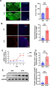
Gradual increase in IGF1 expression in the trigeminal nucleus caudalis (TNC) brain region following repeated intraperitoneal injections of NTG in mice. (A) In the mouse chronic migraine model, there is a significant increase in the number of c-Fos + cells in the trigeminal nucleus caudalis (TNC) brain region. (n = 3 mice/group; compared to saline group, two-tailed independent samples t-test, *p < 0.05, **p < 0.01). (B) In the mouse chronic migraine model, there is a significant increase in CGRP expression in the trigeminal nucleus caudalis (TNC) brain region (n = 5 mice/group; compared to saline group, two-tailed independent samples t-test, *p < 0.05, **p < 0.01). (C) Changes in IGF1 mRNA expression during the induction of chronic migraine in mice (n = 4 mice/group; compared to saline group, two-tailed independent samples t-test, *p < 0.05, **p < 0.01). (D) On the 11th day of the mouse chronic migraine model, there is a significant increase in IGF1 expression in the trigeminal nucleus caudalis (TNC) brain region (n = 5 mice/group; compared to saline group, two-tailed independent samples t-test, *p < 0.05, **p < 0.01). (E) Western blot representative image showing changes in IGF1 expression in the trigeminal nucleus caudalis (TNC) brain region during the mouse chronic migraine model (n = 3 mice/group; two-tailed independent samples t-test, *p < 0.05, **p < 0.01)
Fig. 3
Fig. 3
In the mouse model of chronic migraine, the increase in IGF1 in the trigeminal nucleus caudalis (TNC) brain region primarily originates from autocrine secretion within neuronal cells, with no significant changes observed in IGF1 levels in the liver and blood. (A) Schematic representation of potential sources of IGF1 in the TNC brain region. (B) In the mouse model of chronic migraine, there is no significant change in IGF1 expression in the liver (n = 4 mice/group; compared to saline group, two-tailed independent samples t-test, n.s. p > 0.05). (C) Western blot representative image showing no significant difference in IGF1 expression in circulating blood of mice in the chronic migraine model (n = 3 mice/group; compared to saline group, two-tailed independent samples t-test, n.s. p > 0.05). (D) In the trigeminal nucleus caudalis (TNC) brain region of the mouse chronic migraine model, IGF1 predominantly co-localizes with neurons. Green represents IGF1, and red represents Neun (neuronal marker). (E) In the trigeminal nucleus caudalis (TNC) brain region of the mouse chronic migraine model, IGF1 shows no significant co-localization with microglia. Green represents IGF1, and red represents Iba1 (microglial marker)
Fig. 4
Fig. 4
In the mouse model of chronic migraine, there is an increase in IGF1 receptor phosphorylation in the trigeminal nucleus caudalis (TNC) brain region. Moreover, phosphorylated IGF1r predominantly co-localizes with neurons. (A) Western blot representative image showing the phosphorylation levels of IGF1r in the TNC brain region of the mouse chronic migraine model. (B) Increased phosphorylation of IGF1r in the TNC brain region of mice induced by NTG in the chronic migraine model (n = 4 mice/group; compared to saline group, two-tailed independent samples t-test, *p < 0.05, **P < 0.01). (C) Phosphorylated IGF1r primarily co-localizes with neurons in the mouse chronic migraine model. Green represents phosphorylated IGF1r, and red represents Neun (neuronal marker). (D) Increased number of neurons with phosphorylated IGF1r in the TNC brain region compared to the control group (n = 6 mice/group; compared to saline group, two-tailed independent samples t-test, *p < 0.05, **P < 0.01). (E) In a chronic migraine model of mice, the proportion of neurons with phosphorylated IGF1 receptors in the TNC brain region significantly increased compared to the saline group (n = 5 mice/group; two-tailed independent samples t-test, *P < 0.05, **P < 0.01)
Fig. 5
Fig. 5
Blocking the IGF1/IGF1r pathway alleviates mechanical and thermal hyperalgesia and reduces CGRP expression in the TNC brain region in a mouse model of chronic migraine. (A) Experimental schematic diagram. In the NTG-induced chronic migraine mouse model, mice were intraperitoneally injected with the IGF1 receptor antagonist Picropodophyllin (ppp) at 40 mg/kg two hours before baseline behavioral tests. (B) The IGF1 receptor antagonist attenuated the decreased mechanical withdrawal threshold in the mouse plantar test (n = 5 mice/group; compared to the NTG group, two-tailed independent samples t-test, *p < 0.05, **p < 0.01). (C) The IGF1 receptor antagonist attenuated the decreased mechanical withdrawal threshold around the orbit induced by nitroglycerin in mice (n = 5 mice/group; compared to the NTG group, two-tailed independent samples t-test, *p < 0.05, **p < 0.01). (D) The IGF1 receptor antagonist attenuated the acute mechanical withdrawal threshold decrease in the mouse plantar test induced by nitroglycerin (n = 5 mice/group; compared to the NTG vehicle group, two-tailed independent samples t-test, *p < 0.05, **p < 0.01). (E) The IGF1 receptor antagonist attenuated the decreased thermal withdrawal latency induced by nitroglycerin in mice (n = 5 mice/group; compared to the NTG group, two-tailed independent samples t-test, *p < 0.05, **p < 0.01). (F) The IGF1 receptor antagonist reduced CGRP expression in the TNC brain region induced by nitroglycerin in mice (n = 5 mice/group; compared to the NTG group, two-tailed independent samples t-test, *p < 0.05, **p < 0.01)
Fig. 6
Fig. 6
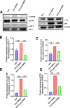
PPP inhibited the overactivation of the mTOR pathway in a chronic migraine mouse model and alleviated neuronal autophagy dysfunction. (A) Representative Western blots. (B) In the chronic migraine mouse model, phosphorylation of IGF1 receptor (IGF1r) is increased in the NTG group. PPP can inhibit this NTG-induced increase in IGF1r phosphorylation (n = 3 mice/group; compared to NTG group, two-tailed independent samples t-test, *p < 0.05, **p < 0.01). (C) In the chronic migraine mouse model, expression of c-Fos is increased in the NTG group. Administration of PPP reduces c-Fos expression in the TNC (n = 3 mice/group; compared to NTG group, two-tailed independent samples t-test, *p < 0.05, **p < 0.01). (D) In the chronic migraine mouse model, phosphorylation of mTOR is increased in the NTG group. PPP can inhibit this NTG-induced increase in mTOR phosphorylation (n = 3 mice/group; compared to NTG group, two-tailed independent samples t-test, *p < 0.05, **p < 0.01). (E) In the chronic migraine mouse model, expression of p62 is increased in the NTG group. PPP can inhibit p62 expression in the NTG model (n = 3 mice/group; compared to NTG group, two-tailed independent samples t-test, *p < 0.05, **p < 0.01)
Fig. 7
Fig. 7
Tail vein injection of IGF1 in mice lowers the facial mechanical withdrawal threshold. An mTOR inhibitor alleviates pain behaviors and ameliorates neuronal autophagy dysfunction. (A) Tail vein injection of IGF1 causes a decrease in the facial mechanical withdrawal threshold in mice, and the autophagy inducer rapamycin partially restores the facial mechanical withdrawal threshold in mice. (B) Representative Western blots. (C) Administration of IGF1 increases phosphorylation of mTOR in the TNC of mice (n = 3 mice/group; compared to NTG group, compared to saline group, two-tailed independent samples t-test, *p < 0.05, **p < 0.01). (D) Administration of IGF1 increases phosphorylation of IGF1 receptor (IGF1r) in the TNC of mice, and rapamycin administration does not affect IGF1 receptor phosphorylation (n = 3 mice/group; compared to IGF1 group, compared to saline group, two-tailed independent samples t-test, *p < 0.05, **p < 0.01). (E) Administration of IGF1 increases c-Fos expression in the TNC of mice, and rapamycin reduces the increase in c-Fos induced by IGF1 (n = 3 mice/group; compared to NTG group, compared to IGF1 group, two-tailed independent samples t-test, *p < 0.05, **p < 0.01). (F) Administration of IGF1 increases p62 expression in the TNC of mice, and rapamycin reduces the increase in p62 induced by IGF1 (n = 3 mice/group; compared to NTG group, compared to IGF1 group, two-tailed independent samples t-test, *p < 0.05, **p < 0.01)

References

    1. Feigin VL, Nichols E, Alam T, Bannick MS, Beghi E, Blake N, Culpepper WJ, Dorsey ER, Elbaz A, Ellenbogen RG et al (2019) Global, regional, and national burden of neurological disorders, 1990–2016: a systematic analysis for the global burden of Disease Study 2016. Lancet Neurol 18:459–480. 10.1016/s1474-4422(18)30499-x - PMC - PubMed
    1. Schwedt TJ (2014) Chronic migraine. BMJ 348:g1416. 10.1136/bmj.g1416 - PubMed
    1. Andreou AP, Edvinsson L (2019) Mechanisms of migraine as a chronic evolutive condition. J Headache Pain 20:117. 10.1186/s10194-019-1066-0 - PMC - PubMed
    1. Dodick DW, Migraine (2018) Lancet 391:1315–1330. 10.1016/s0140-6736(18)30478-1 - PubMed
    1. Latremoliere A, Woolf CJ (2009) Central sensitization: a generator of pain hypersensitivity by central neural plasticity. J Pain 10:895–926. 10.1016/j.jpain.2009.06.012 - PMC - PubMed

MeSH terms