. 2024 Sep 20;30(1):156.
doi: 10.1186/s10020-024-00935-3.

An anti-eCIRP strategy for necrotizing enterocolitis

Affiliations

An anti-eCIRP strategy for necrotizing enterocolitis

Colleen P Nofi et al. Mol Med. .

Abstract

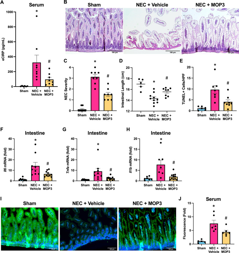
Background: Necrotizing enterocolitis (NEC) is a severe gastrointestinal disease characterized by intestinal inflammation and injury, with high mortality risk. Extracellular cold-inducible RNA-binding protein (eCIRP) is a recently discovered damage-associated molecular pattern that propagates inflammation and tissue injury; however, the role of eCIRP in NEC remains unknown. We hypothesize that eCIRP exacerbates NEC pathogenesis and the novel eCIRP-scavenging peptide, milk fat globule-epidermal growth factor-factor VIII (MFG-E8)-derived oligopeptide 3 (MOP3), attenuates NEC severity, serving as a new therapeutic strategy to treat NEC.

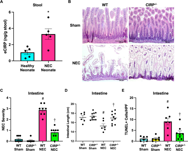
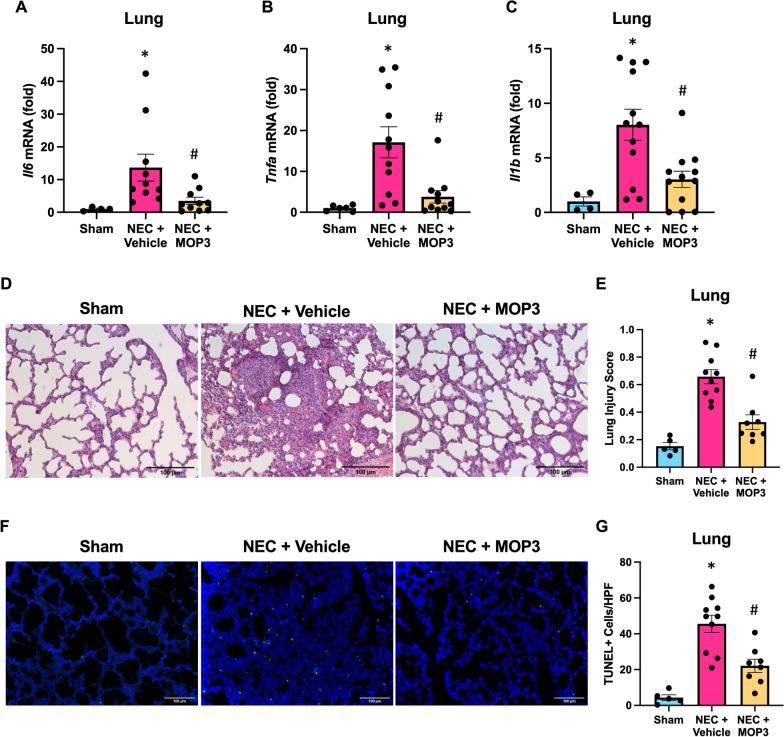
Methods: Stool samples from premature neonates were collected prospectively and eCIRP levels were measured. Wild-type (WT) and CIRP-/- mouse pups were subjected to NEC utilizing a combination of hypoxia and hypercaloric formula orogastric gavage with lipopolysaccharide supplementation. In parallel, WT pups were treated with MOP3 or vehicle. Endpoints including NEC severity, intestinal injury, barrier dysfunction, lung injury, and overall survival were determined.

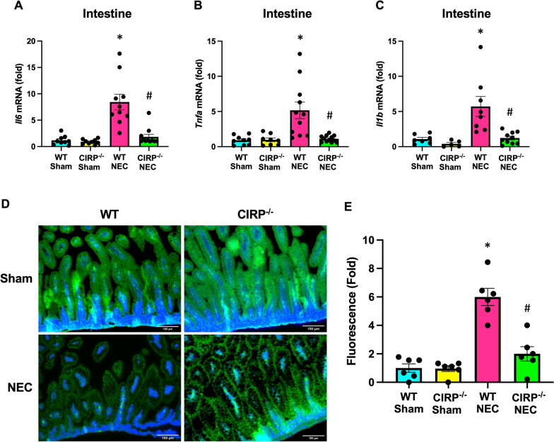
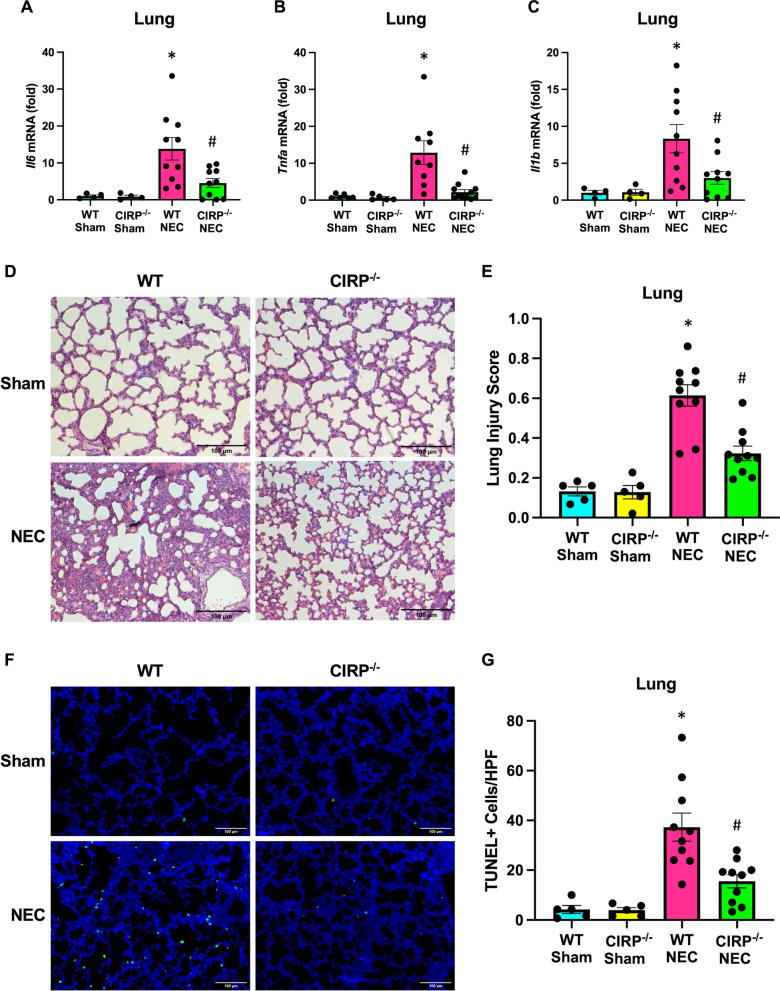
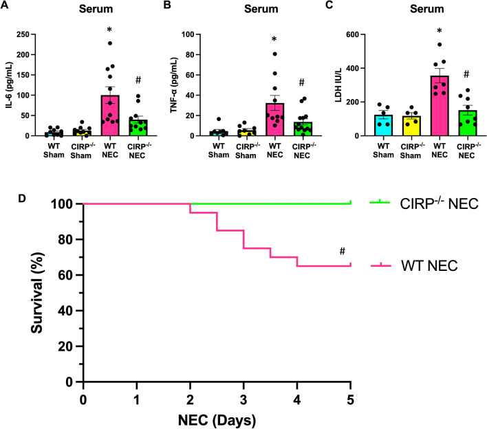
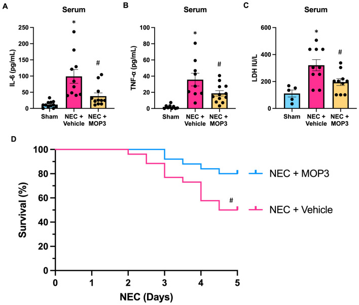
Results: Stool samples from NEC neonates had elevated eCIRP levels compared to healthy age-matched controls (p < 0.05). CIRP-/- pups were significantly protected from NEC severity, intestinal injury, bowel inflammation, intestinal barrier dysfunction, lung injury, and systemic inflammation. NEC survival was 100% for CIRP-/- pups compared to 65% for WT (p < 0.05). MOP3 treatment recapitulated the benefits afforded by CIRP-knockdown, preventing NEC severity, improving inflammatory profiles, and attenuating organ injury. MOP3 treatment improved NEC survival to 80% compared to 50% for vehicle treatment (p < 0.05).

Conclusions: eCIRP exacerbates NEC evidenced by protection with CIRP-deficiency and administration of MOP3, a CIRP-directed therapeutic, in a murine model. Thus, eCIRP is a novel target with human relevance, and MOP3 is a promising treatment for lethal NEC.

Keywords: Inflammation; MFG-E8; MOP3; Necrotizing enterocolitis; eCIRP.


Conflict of interest statement

The authors (CN, MA, and PW) are inventors of the pending PCT application on “A chimeric molecule to treat septic patients”; EFS ID: 47240580; Application No.: 63433789. Other authors report no financial competing interests.

Figures

Fig. 1
Fig. 1
eCIRP exacerbates NEC pathogenesis and intestinal injury. A eCIRP levels detected in the stool of neonates with NEC and age-matched control neonates without NEC. B NEC was induced in WT and CIRP−/− pups and intestines were collected after 4 days of NEC. Age- and litter-matched WT and CIRP−/− sham counterpart intestines were collected for comparison. Representative H&E images of sham and NEC intestines. Original magnification: 200x, Scale bar: 100 μm. C Quantification of NEC severity scoring of H&E intestinal sections. D Quantification of intestinal length from sham and NEC intestines. E Quantification of terminal deoxynucleotidyl transferase dUTP nick end labeling (TUNEL)–positive cells from intestinal sections. All experiments were performed 3 times, and all quantitative data were used for analysis (N = 6–10). Data are expressed as mean ± SEM and compared by One-way ANOVA and Tukey’s method. *p < 0.05 v. healthy neonate, #p < 0.05 vs. sham, p < 0.05 vs. WT NEC
Fig. 2
Fig. 2
eCIRP deficiency protects against intestinal inflammation and barrier dysfunction. NEC was induced in WT and CIRP−/− pups and blood and intestines were collected after 4 days of NEC. Age and litter-matched WT and CIRP−/− sham counterpart blood and intestines were collected for comparison. AC Intestines were frozen and mRNA expression of IL-6, TNFα, and IL-1β was measured by PCR. D Sectioned intestines were stained by immunofluorescence for ZO-1 (green) and nuclear counterstained (blue). Original magnification: 200x, Scale bar: 100 μm. E Pups were administered FITC-dextran (FD4) by orogastric gavage 3-h prior to collection of blood and serum was analyzed for fluorescence intensity (expressed as fold-change from WT sham) as a measure of intestinal permeability. All experiments were performed 3 times, and all quantitative data were used for analysis (N = 6–12). Data are expressed as mean ± SEM and compared by One-way ANOVA and Tukey’s method. *p < 0.05 vs. WT sham, #p < 0.05 vs. WT NEC
Fig. 3
Fig. 3
eCIRP worsens NEC-induced acute lung injury. NEC was induced in WT and CIRP−/− pups and lungs were collected after 4 days of NEC. Age and litter-matched WT and CIRP−/− sham counterpart lungs were collected for comparison. AC Whole lung tissue was frozen and mRNA expression of IL-6, TNFα, and IL-1β were measured by PCR. D Representative H&E images of NEC and sham lungs. Original magnification: 200x, Scale bar: 100 μm. E Quantification of lung injury of representative H&E lung sections by validated lung injury scoring. F Representative terminal deoxynucleotidyl transferase dUTP nick end labeling assay (TUNEL) staining (green) of sham and NEC lungs for apoptotic cells with nuclear counterstain (blue). Original magnification: 200x, Scale bar: 100 μm. G Quantification of TUNEL–positive cells from representative lung sections. All experiments were performed 3 times, and all quantitative data were used for analysis (N = 6–11). Data are expressed as mean ± SEM and compared by One-way ANOVA and Tukey’s method. *p < 0.05 vs. WT sham, #p < 0.05 vs. WT NEC
Fig. 4
Fig. 4
eCIRP deficiency protects against systemic inflammation and mortality in NEC. NEC was induced in WT and CIRP−/− pups and blood was collected after 4 days of NEC. Age and litter-matched WT and CIRP−/− sham counterpart blood was collected for comparison. AB Serum was separated and analyzed for systemic IL-6 and TNFα levels by ELISA. C Serum was analyzed for LDH levels by calorimetric assays. All experiments were performed 3 times, and all quantitative data were used for analysis (N = 6–13). Data are expressed as mean ± SEM and compared by One-way ANOVA and Tukey’s method. D WT and CIRP−/− pups were subjected to NEC for 4 days and monitored for differences in mortality in a 5-day survival study. Overall survival at 5-days was 100% for CIRP−/− pups compared to 65% for WT pups (p = 0.004 by Kaplan Meier and log-rank test, N = 20/group). *p < 0.05 vs. WT sham, #p < 0.05 vs. WT NEC; For survival curve: #p < 0.05 vs. CIRP−/− NEC
Fig. 5
Fig. 5
MOP3 protects against NEC pathogenesis, intestinal injury and inflammation, and barrier dysfunction. NEC was induced in WT pups, and pups were treated with MOP3 (20 μg/g, i.p.) or vehicle (volume equivalent) daily. Intestines were collected after 4 days of NEC. Age and litter-matched sham counterpart intestines were collected for comparison. A eCIRP levels detected in the serum from NEC mice. B Representative H&E images of sham and NEC intestines. Original magnification: 200x, Scale bar: 100 μm. C Quantification of NEC severity scoring of H&E intestinal sections. D Quantification of intestinal length from sham and NEC intestines. E Quantification of terminal deoxynucleotidyl transferase dUTP nick end labeling (TUNEL)–positive cells from intestinal sections. FH Intestines were frozen and mRNA expression of IL-6, TNFα, and IL-1β was measured by PCR. I Sectioned intestines were stained by immunofluorescence for ZO-1 (green) and nuclear counterstained (blue). Original magnification: 200x, Scale bar: 100 μm. J Pups were administered FITC-dextran (FD4) by orogastric gavage 3-h prior to collection of blood and serum was analyzed for fluorescence intensity (expressed as fold-change from sham) as a measure of intestinal permeability. All experiments were performed 3 times, and all quantitative data were used for analysis (N = 6–11). Data are expressed as mean ± SEM and compared by One-way ANOVA and Tukey’s method. *p < 0.05 vs. sham, #p < 0.05 vs. NEC + Vehicle
Fig. 6
Fig. 6
MOP3 attenuates NEC-induced acute lung injury. NEC was induced in WT pups, and pups were treated with MOP3 (20 μg/g, i.p.) or vehicle (volume equivalent) daily. Lungs were collected after 4 days of NEC. Age and litter-matched sham counterpart lungs were collected for comparison. AC Whole lung tissue was frozen and mRNA expression of IL-6, TNFα, and IL-1β were measured by PCR. D Representative H&E images of NEC and sham lungs. Original magnification: 200x, Scale bar: 100 μm. E Quantification of lung injury of representative H&E lung sections by validated lung injury scoring. F Representative terminal deoxynucleotidyl transferase dUTP nick end labeling assay (TUNEL) staining (green) of sham and NEC lungs for apoptotic cells with nuclear counterstain (blue). Original magnification: 200x, Scale bar: 100 μm. G Quantification of TUNEL–positive cells from representative lung sections. All experiments were performed 3 times, and all quantitative data were used for analysis (N = 6–12). Data are expressed as mean ± SEM and compared by One-way ANOVA and Tukey’s method. *p < 0.05 vs. sham, #p < 0.05 vs. NEC + Vehicle
Fig. 7
Fig. 7
MOP3 reduces systemic inflammation and mortality in NEC. NEC was induced in WT pups, and pups were treated with MOP3 (20 μg/kg, i.p.) or vehicle (volume equivalent) daily. Blood was collected after 4 days of NEC. Age and litter-matched sham counterpart blood was collected for comparison. AB Serum was separated and analyzed for systemic IL-6 and TNFα levels by ELISA. C Serum was analyzed for LDH levels by calorimetric assays. All experiments were performed 3 times, and all quantitative data were used for analysis (N = 6–12). Data are expressed as mean ± SEM and compared by One-way ANOVA and Tukey’s method. D Pups were subjected to NEC and treated with MOP3 (20 μg/kg, i.p.) or vehicle (volume equivalent) daily and monitored for differences in mortality in a 5-day survival study. Overall survival at 5-days was 80% for MOP3-treated pups compared to 50% for vehicle-treated pups (p = 0.024 by Kaplan Meier and log-rank test, N = 25–26/group). *p < 0.05 vs. sham, #p < 0.05 vs. NEC + Vehicle; For survival curve: #p vs. NEC + MOP3

Similar articles

Cited by

References

    1. Asaro JA, Khan Z, Brewer M, Klose K, Pesce C, Schanler RJ, et al. Relationship between milk fat globule-epidermal growth factor 8 and intestinal cytokines in infants born preterm. J Pediatr. 2021;230:71–5. - PubMed
    1. Aziz M, Jacob A, Matsuda A, Wang P. milk fat globule-EGF factor 8 expression, function and plausible signal transduction in resolving inflammation. Apoptosis. 2011;16(11):1077–86. - PubMed
    1. Aziz M, Brenner M, Wang P. Extracellular CIRP (eCIRP) and inflammation. J Leukoc Biol. 2019;106(1):133–46. - PMC - PubMed
    1. Aziz M, Prince JM, Wang P. Gut microbiome and necrotizing enterocolitis: understanding the connection to find a cure. Cell Host Microbe. 2022;30(5):612–6. - PubMed
    1. Bazacliu C, Neu J. Necrotizing enterocolitis: long term complications. Curr Pediatr Rev. 2019;15(2):115–24. - PubMed

LinkOut - more resources