. 2024 Sep 20;20(1):419.
doi: 10.1186/s12917-024-04241-2.

Pumpkin seed oil: unveiling its potential in controlling inflammation and pathogenicity during experimental trichinellosis

Affiliations

Pumpkin seed oil: unveiling its potential in controlling inflammation and pathogenicity during experimental trichinellosis

Sara Salah Abdel-Hakeem et al. BMC Vet Res. .

Abstract

Background: This study aimed to investigate the antiparasitic and anti-inflammatory potential of pumpkin seed oil in mice infected with Trichinella spiralis by demonstrating its impact on MMP-9 expression and pathogenesis during the intestinal and muscular phases.

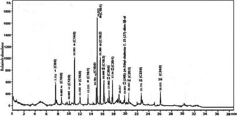
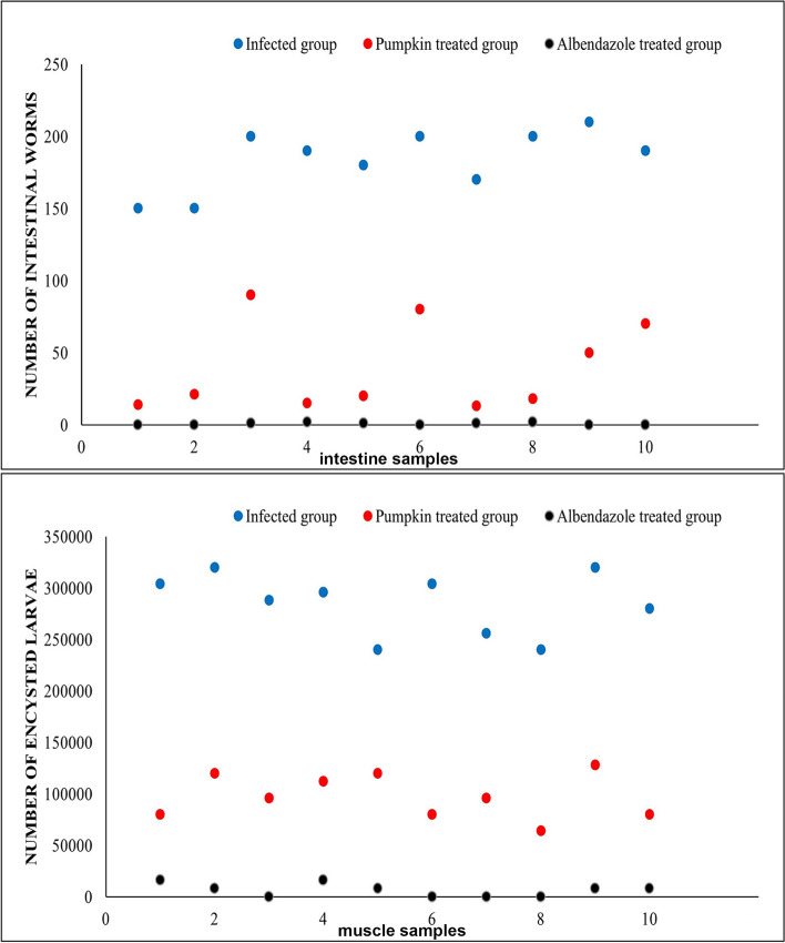
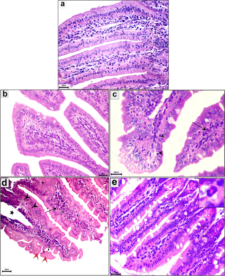
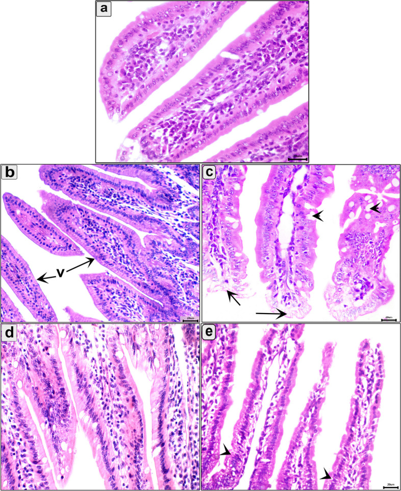
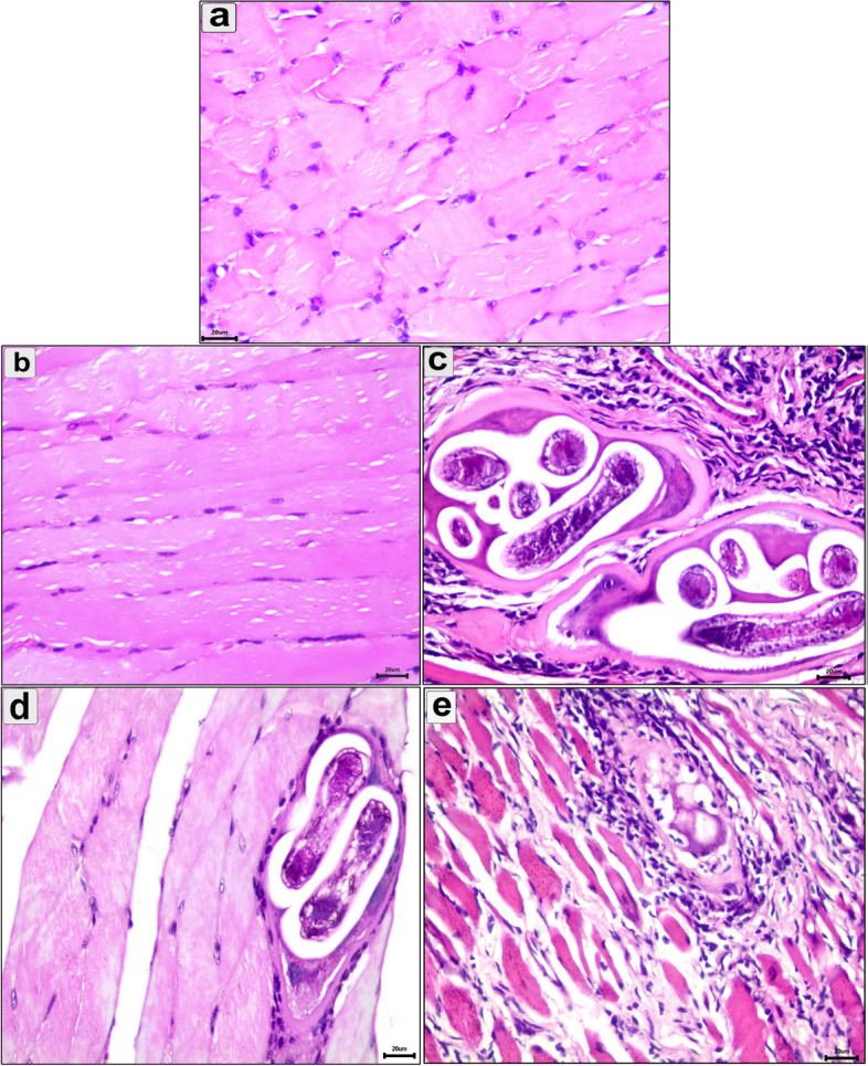
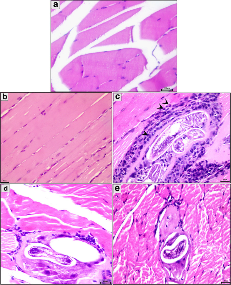
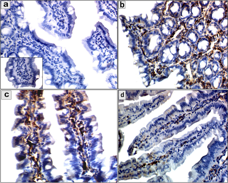
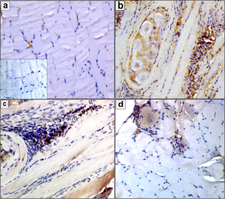
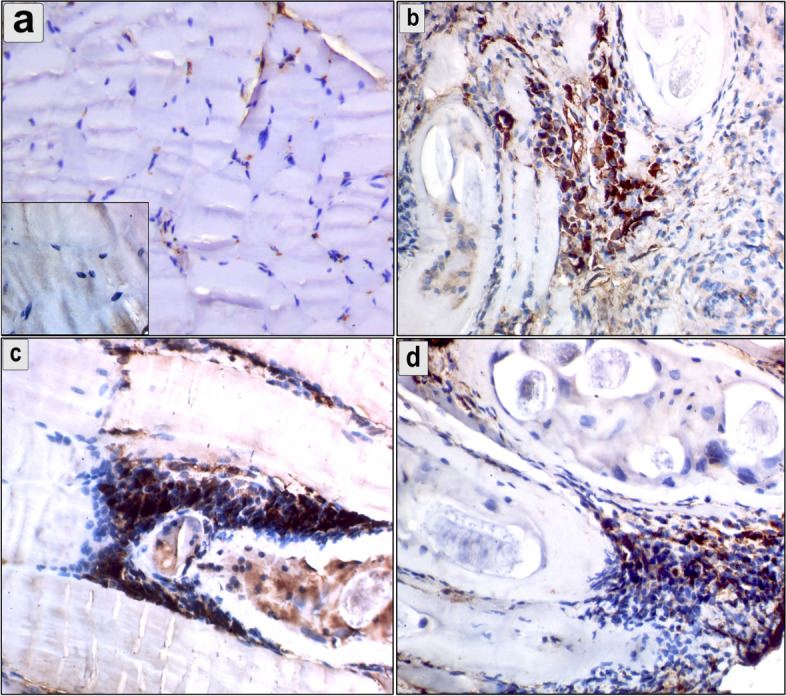
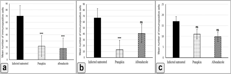
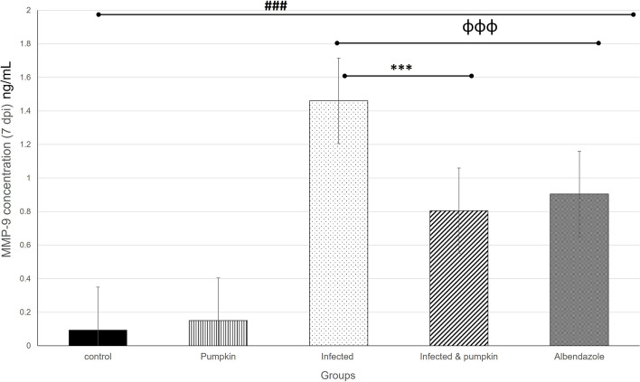
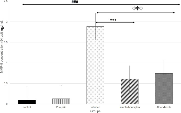
Results: In this study, 100 mice were divided into five groups: an infected group, a pumpkin seed oil-treated group (1.5 mg/kg BW, administered three times per week), an albendazole-treated group, a native control group, and a pumpkin oil control group. Gas chromatography-mass spectrometry analysis of the pumpkin seed oil revealed a broad spectrum of biologically active compounds. The pumpkin seed oil treatment led to a significant reduction in the parasite burden, with a 75% decrease in adult worms and a 66% decrease in encysted larvae. Additionally, the infected animals treated with pumpkin oil exhibited a marked reduction in intestinal inflammation, characterized by a progressive increase in goblet cells. The number of encysted larvae in the diaphragm and muscle tissues was also significantly decreased. Furthermore, pumpkin seed oil treatment significantly reduced MMP-9 levels in both intestinal and muscular tissues, highlighting its potential to attenuate inflammation.

Conclusion: These findings underscore the effectiveness of pumpkin seed oil as anti-inflammatory and antiparasitic agent.

Keywords: Trichinella; Anti-inflammatory; Antiparasitic; MMP-9; Pumpkin seed oil.


Conflict of interest statement

The authors declare no competing interests.

Figures

Fig. 1
Fig. 1
GC-Mass histogram shows the main sixteen phytochemical constituents in the pumpkin seed oil used in the current evaluation
Fig. 2
Fig. 2
Dot plot shows a marked reduction in mean number of the adult worms (a) and encysted larvae (b) in the pumpkin and albendazole-treated groups
Fig. 3
Fig. 3
Photomicrograph of the intestine in the different groups in the enteral phase showing: a Normal structure of the intestinal mucosa; b uninfected pumpkin-treated intestine showing normal villi with healthy enterocytes; c Transverse section (T.s) in the intestine of the infected untreated group showing the parasitic enteritis characterized by shedding of the epithelium high inflammatory reaction in the core of the villi (arrow) and mononuclear cells between enterocytes (arrowhead); d T.s in the intestine of the pumpkin-treated group showing less normal intestinal mucosa, hyperplasia in some enterocytes (arrowhead), desquamation of the epithelium in tips of villi (red arrowhead), and mild inflammatory reaction in the core of villi (arrow); e T.s in the intestine of the albendazole-treated group showing normal intestinal villi with high number of goblet cells and eosinophils infiltration (small box). Stain H&E (400 ×)
Fig. 4
Fig. 4
Photomicrograph of the intestine in the different groups in the parenteral phase showing: a Normal structure of the intestinal mucosa; b T.s in the intestine of uninfected pumpkin-treated showing normal villi (v) with healthy enterocytes; c T.s in the intestine of infected untreated group showing vacuolar degeneration in the enterocytes at the tips of villi (arroe) and increase the number of goblet cell (arrowhead); d T.s in the intestine of pumpkin-treated group showing normal architecture of intestinal mucosa; e T.s in the intestine of albendazole-treated group showing necrobiotic changes and vacuolar degeneration of enterocytes (arrowhead). Stain H&E (400 ×)
Fig. 5
Fig. 5
Photomicrograph of the diaphragm in the different groups showing: a Normal diaphragm consists of groups of striated muscle fibers; b The diaphragm of uninfected pumpkin-treated group showing normal muscle fibers; c The diaphragm of infected untreated group showing multiple well-developed cysts containing many larvae surrounded by connective tissue capsule; d The diaphragm of pumpkin-treated group showing marked reduction in the number and size of encysted larvae with normal architecture of the muscle fibers; e The diaphragm of albendazole-treated group showing degeneration larvae and muscle fibers with marked inflammatory reaction. Stain H&E (400 ×)
Fig. 6
Fig. 6
Photomicrograph of the muscle in the different groups showing: a Normal structure of muscle consists of bands of muscle fibers; b Muscle section of the uninfected pumpkin-treated group showing health muscle fibers with mild inflammatory reaction; c Muscle section of the infected untreated group showing multiple larval cysts surrounded by severe inflammatory cells, particularly eosinophils (arrowhead); d Muscle section of the pumpkin-treated group showing a degenerative encysted larvae with mild inflammatory reaction; e Muscle section of the albendazole-treated group showing minute encysted larvae with mild degeneration in the muscle fibers. Stain H&E (400 ×)
Fig. 7
Fig. 7
An immunohistochemical staining of MMP-9 the intestine demonstrates the anti-inflammatory effects of pumpkin oil and albendazole (a) The negative reaction in the uninfected pumpkin-treated group (negative control in the small box); b Severe reaction of MMP-9 positive cells in the infected untreated group were distributed in the mucosa and extending to the submucosa; c Marked downregulation in the MMP-9 expression in the pumpkin-treated group and the albendazole-treated group (d)
Fig. 8
Fig. 8
An immunohistochemical staining of MMP-9 in the diaphragm demonstrates the anti-inflammatory effects of pumpkin seed oil and albendazole (a) The negative reaction in the uninfected pumpkin-treated group (negative control in the small box); b Cytoplasmic reaction of MMP-9 positive cells were distributed around encysted larvae and between muscle bands in the infected untreated group; c Marked downregulation in the expression of MMP-9 around the encysted larvae in the pumpkin-treated group and the albendazole-treated group (d)
Fig. 9
Fig. 9
An immunohistochemical staining of MMP-9 in the muscle demonstrates the anti-inflammatory effects of pumpkin and albendazole. a The negative reaction in the uninfected pumpkin-treated group (negative control in the small box); b Cytoplasmic reaction of MMP-9 positive cells in the infected untreated group were distributed between muscle fibers and around the encysted larvae; c non-significant reduction in the MMP-9 expression was observed around the encysted larvae and between muscle bands in the pumpkin-treated group and the albendazole-treated group (d)
Fig. 10
Fig. 10
Histogram showing the mean number of MMP-9 positive cells in the intestine (a), diaphragm (b), and muscle (c) of the different groups. Differences were highly significant at ***P ≤ 0.0001, ns, non-significant
Fig. 11
Fig. 11
Histogram showing the quantitative level of MMP-9 in the enteral phase of the different groups. A highly significant increase in the level of MMP-9 was observed between uninfected and infected groups. A significant reduction in the treated groups compared to the infected untreated group. Differences were highly significant at P ≤ 0.0001
Fig. 12
Fig. 12
Histogram showing the quantitative level of MMP-9 in the parenteral phase of the different groups. A highly significant increase in the level of MMP-9 was observed between uninfected and infected groups. A significant reduction in the treated groups compared to the infected untreated group. Differences were highly significant at P ≤ 0.0001

Similar articles

Cited by

References

    1. Liu X, Song Y, Jiang N, Wang J, Tang B, Lu H, Peng S, Chang Z, Tang Y, Yin J. Global gene expression analysis of the zoonotic parasite Trichinella spiralis revealed novel genes in host parasite interaction. 2012. - PMC - PubMed
    1. Yadav AK. Temjenmongla: Efficacy of Lasia spinosa leaf extract in treating mice infected with Trichinella spiralis. Parasitol Res. 2012;110:493–8. - PubMed
    1. Gottstein B, Pozio E, Nöckler K. Epidemiology, diagnosis, treatment, and control of trichinellosis. Clin Microbiol Rev. 2009;22(1):127–45. - PMC - PubMed
    1. Nassef NE, Moharm IM, Atia AF, Brakat RM, Abou Hussien NM, Shamseldeen A. Therapeutic efficacy of chitosan nanoparticles loaded with albendazole on parenteral phase of experimental Trichinellosis. J Egypt Soc Parasitol. 2019;49(2):301–11.
    1. Speich B, Ali SM, Ame SM, Bogoch II, Alles R, Huwyler J, Albonico M, Hattendorf J, Utzinger J, Keiser J. Efficacy and safety of albendazole plus ivermectin, albendazole plus mebendazole, albendazole plus oxantel pamoate, and mebendazole alone against Trichuris trichiura and concomitant soil-transmitted helminth infections: a four-arm, randomised controlled trial. Lancet Infect Dis. 2015;15(3):277–84. - PubMed

LinkOut - more resources