. 2024 Sep 20;24(1):1173.
doi: 10.1186/s12885-024-12948-6.

Type 2-like polarization and elevated CXCL4 secretion of monocyte derived macrophages upon internalization of plasma-derived exosomes from head and neck cancer patients

Affiliations

Type 2-like polarization and elevated CXCL4 secretion of monocyte derived macrophages upon internalization of plasma-derived exosomes from head and neck cancer patients

Marie-Nicole Theodoraki et al. BMC Cancer. .

Abstract

Background: Exosomes are closely associated with different aspects of tumor-progression in patients with head and neck squamous cell carcinoma (HNSCC), such as angiogenesis or immune regulation. As extracellular vesicles they are involved in the intercellular communication by transferring their cargo such as proteins and nucleic acids from one cell to another. However, the influence of tumor related plasma-derived exosomes on the polarization and characteristics of monocyte derived macrophages is not fully understood.

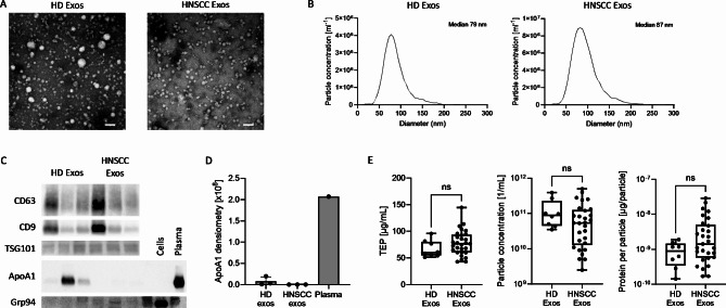
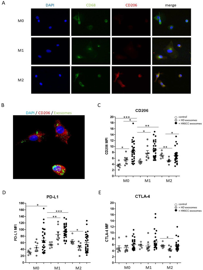
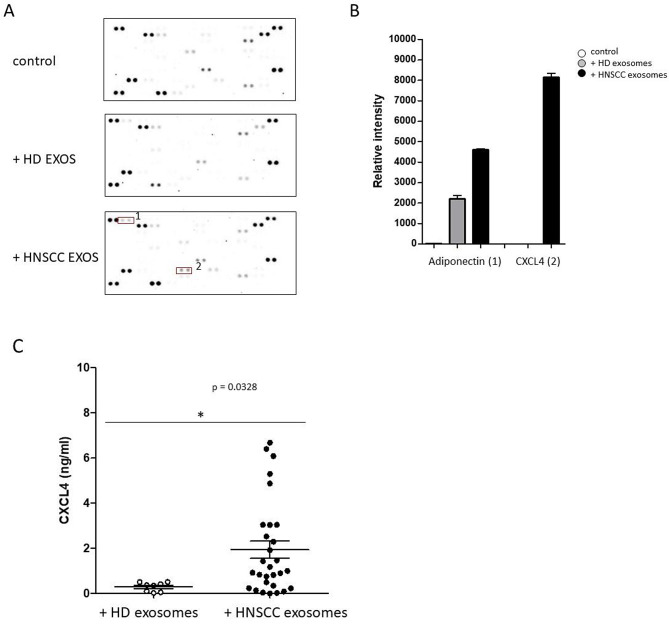
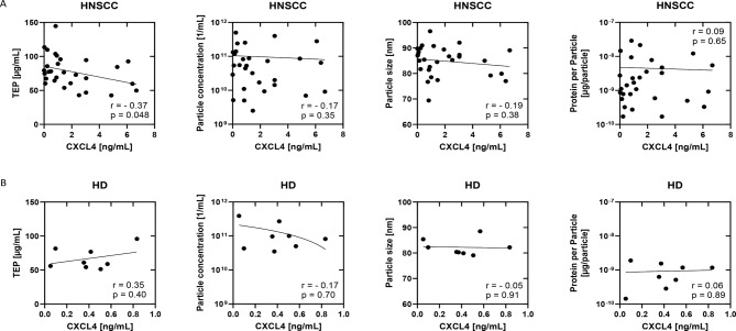
Methods: Exosomes were isolated from plasma samples of healthy donors (HD) and HNSCC patients and further evaluated with regard to morphology, size and protein composition via transmission electron microscopy, nanoparticle tracking, western blot analysis and cytokine assays. Differentiation and characteristics of monocyte derived macrophages upon exosome internalization were analyzed using flow cytometry and fluorescence microscopy. Macrophage cytokine secretion patterns were analyzed by human cytokine antibody arrays and ELISA measurements.

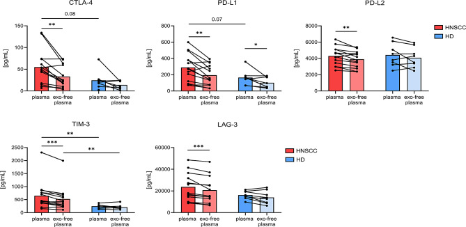
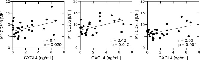
Results: Our data revealed elevated overall plasma levels of CTLA-4, PD-L1, and TIM-3 as well as elevated exosome-associated CTLA-4, PD-L2, TIM-3, and LAG-3 levels in HNSCC patients compared to HD. Furthermore, we observed a significant type 2-like polarization and elevated CXCL4 secretion of monocyte derived macrophages upon internalization of plasma-derived exosomes from HNSCC patients, which could be visualized by fluorescence microcopy of membrane stained exosomes.

Conclusions: The study provides new insights regarding exosome driven pro-tumorigenic immune regulation in the circulation of patients with head and neck cancer and could help to better understand the individual immunologic situation.

Keywords: CD206; CTLA-4; CXCL4; Exosomes; HNSCC; Liquid biomarker; Macrophages; PD-L1.


Conflict of interest statement

The authors declare no competing interests.

Figures

Fig. 1
Fig. 1
Characterization of plasma exosomes. (A) Representative transmission electron microscopy images and (B) size distribution profiles of exosomes from plasma of a healthy donor (HD) and a patient with head and neck squamous cell carcinoma (HNSCC). Scale bar = 100 nm. (C) Representative western blot images from plasma-derived exosomes. (D) Quantification of ApoA1 band intensity from (C). (E) Comparison of total exosome protein (TEP), particle concentration and protein per particle between n = 8 HD and n = 29 HNSCC patients. Box-and-whiskers show median, 25th and 75th quartiles and range. ns = not significant, as determined by Mann-Whitney test
Fig. 2
Fig. 2
Comparison of immune modulators in plasma before and after depletion of exosomes. Levels of CTLA-4, PD-L1, PD-L2, TIM-3 and LAG-3 were measured with Milliplex Multiplex Assay in plasma of HNSCC patients (n = 14) and HD (n = 8) before (plasma) and after (exo-free plasma) depletion of exosomes by ultracentrifugation. Differences in plasma before and after exosome depletion were compared by paired Wilcoxon test, differences between HNSCC patients and HD by Mann-Whitney test
Fig. 3
Fig. 3
Macrophage polarization of THP-1 monocytes in response to plasma derived exosomes from HDs and HNSCC patients, compared to the control (absence of exosomes). (A) Polarization of THP-1 monocytes into type 1-like macrophages (induced by 20 ng/ml IFN, 10 pg/ml LPS) and type 2-like macrophages (induced by 20 ng/ml IL-4, 20 ng/ml IL-13) compared to non-polarized (M0) macrophages via immunohistochemical staining of M2-like macrophage marker CD206 and pan macrophage marker CD68. Blue fluorescence shows DAPI stained nuclei, green fluorescence shows FITC-anti-CD68 staining and red fluorescence shows dsRed-anti-CD206 staining. (B) Representative exosomal internalization by monocyte derived macrophages was visualized by FITC-fluorescence staining of the exosomal membrane (green). Blue fluorescence shows DAPI stained nuclei and red fluorescence shows dsRed-anti-CD206 staining. (C-E) Expression of M2-like macrophage marker CD206 (C), immune checkpoint molecule PD-L1 (D) and CTLA-4 (D) under M0 and M1 and M2 culturing conditions in response to both HD and HNSCC exosomes. *: p < 0.05; **: p < 0.01; ***: p < 0.001. MFI: mean fluorescence intensity
Fig. 4
Fig. 4
Cytokine screening upon internalization of plasma derived exosomes. (A) Raw images of cytokine arrays of culture supernatants from monocyte derived macrophages upon internalization of plasma derived exosomes from HDs and HNSCC patients. Numbers indicate increased densities of bands of certain cytokines in response to HNSCC exosomes (1: Adiponectin; 2: CXCL4). (B) Semiquantitative analysis was performed by measuring the density of the dots and revealed differential secretion patterns of different cytokines. (C) Quantification of CXCL4 supernatant concentrations (ng/ml) by ELISA measurements revealed significantly increased concentrations of chemokine CXCL4 in response to plasma exosomes from HNSCC patients compared to HDs. *: p < 0.05
Fig. 5
Fig. 5
Correlation analysis between secreted CXCL4 (ng/ml) and expression levels of M2-like macrophage marker CD206 on monocyte derived M0/M1/M2 macrophages upon HNSCC exosome internalization. The correlation coefficient (r) and p values are given for each correlation. p < 0.05 was considered as significant. MFI: mean fluorescence intensity
Fig. 6
Fig. 6
Correlation of exosome characteristics and CXCL4 in HNSCC patients. Scatter plots of correlation analysis of (A) n = 29 HNSCC and (B) n = 8 HD between CXCL4 levels and TEP, particle concentration, particle size or protein per particle. Dependent on the data distribution, Spearman or Pearson correlation analysis was applied. p < 0.05 was considered as significant

References

    1. Zhang P, Li S, Zhang T, Cui F, Shi JH, Zhao F, et al. Characterization of Molecular subtypes in Head and Neck squamous cell carcinoma with distinct prognosis and treatment responsiveness. Front cell Dev Biology. 2021;9:711348. - PMC - PubMed
    1. Sung H, Ferlay J, Siegel RL, Laversanne M, Soerjomataram I, Jemal A, et al. Global Cancer statistics 2020: GLOBOCAN estimates of incidence and Mortality Worldwide for 36 cancers in 185 countries. Cancer J Clin. 2021;71:209–49. - PubMed
    1. Califf RM. Biomarker definitions and their applications. Experimental Biology Med. 2018;243:213–21. - PMC - PubMed
    1. Alsahafi E, Begg K, Amelio I, Raulf N, Lucarelli P, Sauter T, et al. Clinical update on head and neck cancer: molecular biology and ongoing challenges. Cell Death Dis. 2019;10:540. - PMC - PubMed
    1. Canning M, Guo G, Yu M, Myint C, Groves MW, Byrd JK, et al. Heterogeneity of the Head and Neck squamous cell Carcinoma Immune Landscape and its impact on Immunotherapy. Front cell Dev Biology. 2019;7:52. - PMC - PubMed

MeSH terms