. 2024 Sep 20;15(1):316.
doi: 10.1186/s13287-024-03889-9.

MSC-extracellular vesicle microRNAs target host cell-entry receptors in COVID-19: in silico modeling for in vivo validation

Affiliations

MSC-extracellular vesicle microRNAs target host cell-entry receptors in COVID-19: in silico modeling for in vivo validation

Hajer A Al Saihati et al. Stem Cell Res Ther. .

Erratum in

Abstract

Background: Coronavirus disease 2019 (COVID-19) has created a global pandemic with significant morbidity and mortality. SARS-CoV-2 primarily infects the lungs and is associated with various organ complications. Therapeutic approaches to combat COVID-19, including convalescent plasma and vaccination, have been developed. However, the high mutation rate of SARS-CoV-2 and its ability to inhibit host T-cell activity pose challenges for effective treatment. Mesenchymal stem cells (MSCs) and their extracellular vesicles (MSCs-EVs) have shown promise in COVID-19 therapy because of their immunomodulatory and regenerative properties. MicroRNAs (miRNAs) play crucial regulatory roles in various biological processes and can be manipulated for therapeutic purposes.

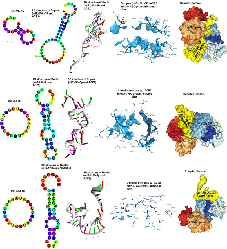
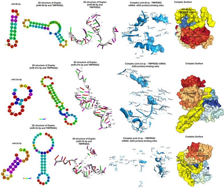
Objective: We aimed to investigate the role of lyophilized MSC-EVs and their microRNAs in targeting the receptors involved in SARS-CoV-2 entry into host cells as a strategy to limit infection. In silico microRNA prediction, structural predictions of the microRNA-mRNA duplex, and molecular docking with the Argonaut protein were performed.

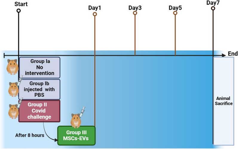
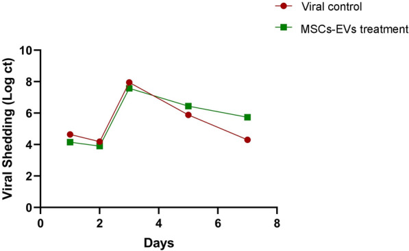
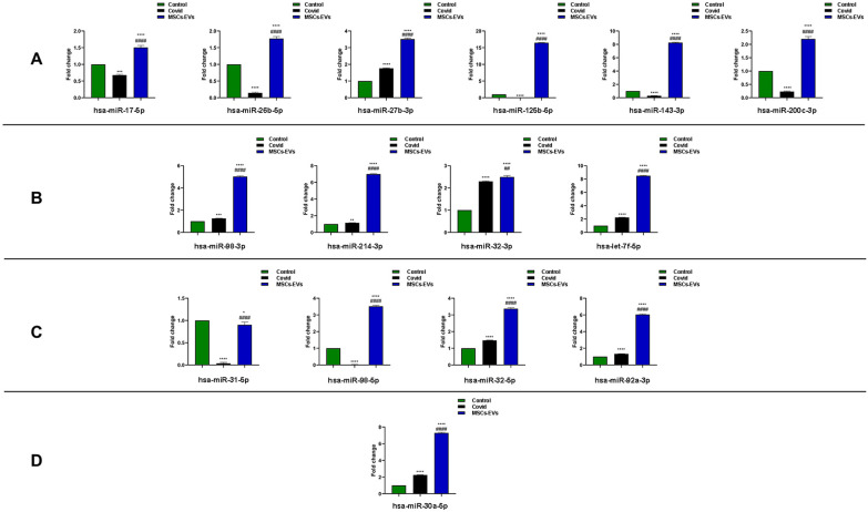
Methods: Male Syrian hamsters infected with SARS-CoV-2 were treated with human Wharton's jelly-derived Mesenchymal Stem cell-derived lyophilized exosomes (Bioluga Company)via intraperitoneal injection, and viral shedding was assessed. The potential therapeutic effects of MSCs-EVs were measured via histopathology of lung tissues and PCR for microRNAs.

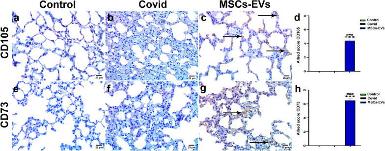
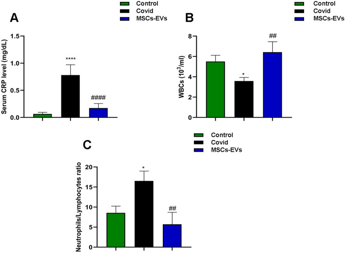
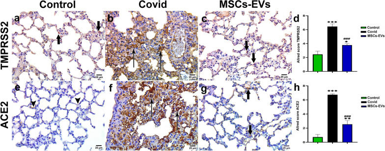
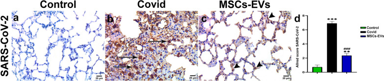
Results: The results revealed strong binding potential between miRNA‒mRNA duplexes and the AGO protein via molecular docking. MSCs-EVs reduced inflammation markers and normalized blood indices via the suppression of viral entry by regulating ACE2 and TMPRSS2 expression. MSCs-EVs alleviated histopathological aberrations. They improved lung histology and reduced collagen fiber deposition in infected lungs.

Conclusion: We demonstrated that MSCs-EVs are a potential therapeutic option for treating COVID-19 by preventing viral entry into host cells.

Keywords: COVID-19; MSC–EVs; MicroRNAs; Molecular docking; Receptors for viral entry.


Conflict of interest statement

Authors declare that they have no known competing financial interests or personal relationships that could have appeared to influence the work reported in this paper.

Figures

Fig. 1
Fig. 1
Timeline of the experimental design
Fig. 2
Fig. 2
2D structural models of miRNAs) miR-200c-3p, miR-26b-5p and miR-125b-5p (; 2D and 3D structural models of microRNAs duplexes with mRNA of ACE2 gene; Binding sites interactions between amino acid residues of Argonaut protein and microRNAs- ACE2 duplexes and surface shape of argonaut silencing complexes
Fig. 3
Fig. 3
2D structural models of miR-17-5p, miR-27b-3p and miR-143-3p; 2D and 3D structural models of different microRNAs duplexes with mRNA of ACE2 gene; Binding sites interactions between amino acid residues of Argonaut protein and microRNAs- ACE2 duplexes and surface shape of argonaut silencing complexes
Fig. 4
Fig. 4
2D structural models of miR-98-5p, miR-214-3p and miR-32-3p; 2D and 3D structural models of different microRNAs (miR-98-5p, miR-214-3p and miR-32-3p) duplexes with mRNA of TMPRESS2 gene; Binding sites interactions between amino acid residues of Argonaut protein and microRNAs- TMPRESS2 duplexes and surface shape of argonaut silencing complexes
Fig. 5
Fig. 5
2D structural models of miR-98-3p, miR-92a-3p and miR-31-5p; 2D and 3D structural models of miR-98-3p with mRNA of ITGA3 gene and miR-92a-3p, miR-31-5p with mRNA of ITGA5 gene; Binding sites interactions between amino acid residues of Argonaut protein and microRNAs- ITGA duplexes and surface shape of argonaut silencing complexes
Fig. 6
Fig. 6
2D structural models of miR-32-5p, miR-30a-5p and let-7f-5p; 2D and 3D structural models of miR-32-5p with mRNA of ITGAV, miR-30a-5p with mRNA of CTSL1, let-7f-5p with mRNA of TMPRESS2 gene; Binding sites interactions between amino acid residues of Argonaut protein and microRNAs- ITGAV, CTSL1 and TMPRESS2 duplexes and surface shape of argonaut silencing complexes
Fig. 7
Fig. 7
Viral shedding is unaltered following exosome treatment. Viral shedding from infected and infected + MSCs–EVs was determined at days 0, 2, 4, 6, and 8. The x-axis indicates time (days), and y-axis indicates Viral Shedding (log ct)
Fig. 8
Fig. 8
Immunohistochemical reaction for MSCs–EVs localization in lung tissues. a control group: negative immune reaction for CD105, b covid group: negative immune reaction for CD105, c MSCs–EVs group: Strong immune reaction for CD105 (arrow). d Allred score for CD105 expressed as mean ± SEM.***p < 0.001 versus the control group, ###p < 0.001 versus the covid group. e control group: negative immune reaction for CD73, f covid group: negative immune reaction for CD73, g MSCs–EVs group: Strong immune reaction for CD73 (arrow). h) Allred score for CD73 expressed as mean ± SEM.***p < 0.001 versus the control group, ###p < 0.001 versus the covid group
Fig. 9
Fig. 9
MSC–EVs administration reduces covid induced inflammatory responses. a Serum CRP; b serum WBCs; c Serum neutrophil /lymphocyte. Data are expressed as mean ± SEM. *p < 0.05 versus the control group, ****p < 0.0001 versus the control group, ##p < 0.01 versus the covid group, ####p < 0.0001 versus the covid group
Fig. 10
Fig. 10
MSC–EVs and their role in miRNA expression regulation. a Fold change of ACE2 receptor interacting microRNAs miR-17-5p, miR-26b-5p, miR-27b-3p, miR-125b-5p, miR-143-3p and miR-200c-3p in control, covid and MSC–EVs groups.; b Fold change of TMPRESS2 receptor interacting microRNAs miR-98-5p, miR-214-3p, miR-32-3p and let-7f-5p in control, covid and MSC–EVs groups. c Fold change of ITGA receptors interacting microRNAs miR-31-5p, miR-98-5p, miR-32-5p and miR-92a-3p in control, covid-infected and MSC–EVs groups.; d Fold change of CTSL1 receptor interacting microRNA miR-30a-5p in control, covid and MSC–EVs groups. Data are expressed as mean ± SEM. *p < 0.05 versus the control group, **p < 0.01 versus the control group, ***p < 0.001 versus the control group, ****p < 0.0001 versus the control group, #p < 0.05 versus the covid group, ##p < 0.01 versus the covid group, ###p < 0.001 versus the covid group, ####p < 0.0001 versus the covid group
Fig. 11
Fig. 11
Representative photomicrographs of lung immune stained sections for ac TMPRSS2 expression a Control group: moderate immune expression (bold arrow) b Covid group: intense brown granular cytoplasmic positivity (arrow) in the alveolar cells c) MSCs–EVs group: mild to moderate brown granular cytoplasmic positivity (bold arrow) in the alveolar cells. ef ACE2 expression e Control group: mild immunoreactivity in some alveolar cells. f Covid group: intense immunoreactivity in a large number of cells. g MSCs–EVs group: moderate reaction in the alveolar cells. d and h Allred score for TMPRSS2 and ACE2 expression. Data are expressed as mean ± SEM. *p < 0.05 versus the control group, **p < 0.01 versus the control group, ***p < 0.001 versus the control group, ###p < 0.001 versus covid group. Allred index (0–1 = negative, 2–3 = mild, 4–6 = moderate, and 7–8 = strongly positive)
Fig. 12
Fig. 12
Representative photomicrographs from SARS-CoV-2 immune stained lung sections showing a Control group: negative immune reactivity b Covid group: intense brown granular cytoplasmic immune reactivity (arrow) in the alveolar epithelium c MSCs–EVs group: mild to moderate brown granular cytoplasmic positivity (arrowhead) in the alveolar epithelium. d Allred score for SARS-CoV-2 immune expression. Data are expressed as mean ± SEM. **p < 0.01 versus the control group, ***p < 0.001 versus the control group, ###p < 0.001 versus the covid group,####p < 0.0001 versus the covid group. Allred index (0–1 = negative, 2–3 = mild, 4–6 = moderate, and 7–8 = strongly positive)
Fig. 13
Fig. 13
Representative photomicrographs from (ac) hematoxylin and eosin and (eg) Masson trichrome stained lung sections. H&E: a Control group: patent alveolar lumens and thin interalveolar septa (double arrow) lined by squamous type I pneumocytes (arrowhead) and large cuboidal type II pneumocytes (short arrow). An alveolar capillary (c) is also seen. b Covid group: Lung consolidation with diffuse thickening of the interalveolar septa, abundant inflammatory cells (if), collapsed lumens (arrow), intra alveolar hemorrhage (asterisk) and congested blood vessels (bv). c MSCs–EVs group: with predominantly patent alveolar lumens and thin interalveolar septa (arrow) and residual thickening of the interalveolar septa with inflammatory cell infiltration (if) and congested blood vessels (bv). d Assessment of lung with the lung injury scoring system. Data are expressed as mean ± SEM.***p < 0.001 versus the control group, ###p < 0.001 versus the covid group. Masson trichrome: e Control group: fine collagen fibers in the interalveolar septa and surrounding the bronchioles and blood vessels. f Covid group: abundant, intensely staining collagen fibers in the thickened interalveolar septa (arrow) and surrounding the bronchioles (double arrow). g MSCs–EVs group: fine, intensely stained collagen fibers in the interalveolar septa (arrow) and around the blood vessels. h Mean area percentage of collagen fibers in the different experimental groups. Data are expressed as mean ± SEM. ***p < 0.001 versus the control group, ###p < 0.001 versus the covid group

References

    1. Cascella M, Rajnik M, Aleem A, Dulebohn SC, Di Napoli R. Features, evaluation, and treatment of coronavirus (COVID-19). Statpearls [internet]. StatPearls publishing; 2023. - PubMed
    1. Cucinotta D, Vanelli M. WHO declares COVID-19 a pandemic. Acta Bio med Atenei Parm Mattioli. 1885;2020(91):157. - PMC - PubMed
    1. Mohammed MA. Fighting cytokine storm and immunomodulatory deficiency: by using natural products therapy up to now. Front Pharmacol. 2023;14:1111329. - PMC - PubMed
    1. Ali FEM, Mohammedsaleh ZM, Ali MM, Ghogar OM. Impact of cytokine storm and systemic inflammation on liver impairment patients infected by SARS-CoV-2: prospective therapeutic challenges. World J Gastroenterol. 2021;27:1531. - PMC - PubMed
    1. Hoffmann M, Kleine-Weber H, Schroeder S, Krüger N, Herrler T, Erichsen S, et al. SARS-CoV-2 cell entry depends on ACE2 and TMPRSS2 and is blocked by a clinically proven protease inhibitor. Cell. 2020;181:271–80. - PMC - PubMed

MeSH terms

LinkOut - more resources