

Surgical management for the case of scrotal myiasis in a 7-day-old neonate: a case report
- PMID: 39304966
- PMCID: PMC11415981
- DOI: 10.1186/s13256-024-04759-x
Surgical management for the case of scrotal myiasis in a 7-day-old neonate: a case report
Abstract
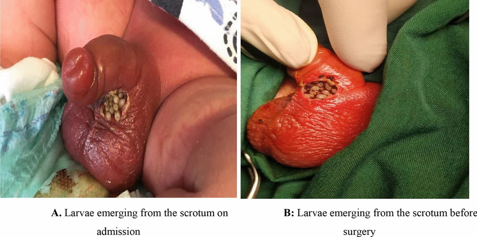
Introduction: Neonatal myiasis is a rare condition, with few reports available on the subject. Surgical management is recommended in some cases. In this study, we present the case of a 7-day-old male neonate with larvae in his scrotum who underwent surgery.
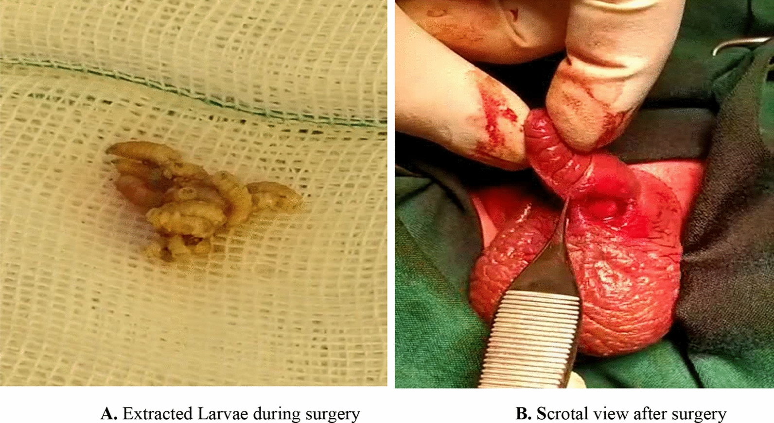
Case presentation: A full-term 7-day-old male infant (Aryan race) was referred to a children's hospital. On the sixth day after birth, three 3-4 mm long larvae crawled out from his scrotum, with the number increasing over time. He was given intravenous antibiotics and topical mupirocin to combat secondary infections. The surgical treatment involved two steps: first, the larvae were extracted, and then the infection site was washed with betadine and hydrogen peroxide to help remove any possible remaining larvae.
Conclusion: Scrotal myiasis is a rare disease that occurs in infants and requires immediate treatment. Surgical treatment is effective in removing dead or decaying larvae from a deep-seated location and washing the infection site to prevent secondary infection.
Keywords: Iran; Myiasis; Neonate; Scrotum; Surgery.
© 2024. The Author(s).
Conflict of interest statement
The authors declare no other conflicts of interest.
Figures
References
-
- Sykes JE, Merkel L, Little SE. Myiasis. In: Greene CE, editor. Greene’s infectious diseases of the dog and cat. Philadelphia: WB Saunders; 2021. p. 1347–58.
-
- Jokar A, Sharififard M, Jahanifard E. Prevalence of human myiasis and its epidemiological aspects in Iran from 2013 to 2020: a review study. J Prev Med. 2022;9(2):102–15.
-
- Cetinkaya M, Ozkan H, Köksal N, Coşkun SZ, Hacimustafaoğlu M, Girişgin O. Neonatal myiasis: a case report. Turk J Pediatr. 2008;50(6):581–4. - PubMed
Publication types
MeSH terms
Substances
LinkOut - more resources
Full Text Sources
