

Novel KIF26A variants associated with pediatric intestinal pseudo-obstruction (PIPO) and brain developmental defects
- PMID: 39305096
- PMCID: PMC11608842
- DOI: 10.1111/cge.14621
Novel KIF26A variants associated with pediatric intestinal pseudo-obstruction (PIPO) and brain developmental defects
Abstract
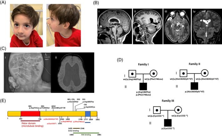
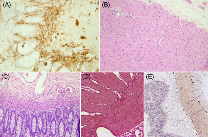
Pediatric intestinal pseudo-obstruction (PIPO) is a rare congenital disorder of the enteric nervous system with distal colon aganglionosis potentially leading to intestinal obstruction. Recently, biallelic variants in KIF26A, encoding a crucial motor protein for the migration and differentiation of enteric neural crest cells, have been associated with a neurodevelopmental condition featuring cortical defects and PIPO-like features, though in absence of aganglionosis. So far, only 10 patients have been reported. In this study, we investigated three subjects with congenital hydrocephalus, neurodevelopmental impairment, and intestinal obstruction megacolon syndrome. Brain MRI revealed malformations within cortical dysplasia spectrum, including polymicrogyria and heterotopia. Pathology study of the intestine revealed aganglionosis and elevated acetylcholinesterase activity in parasympathetic nerve fibers. Through trio-exome sequencing (ES), we detected four novel biallelic KIF26A variants, including two missense changes (#1) and two distinct homozygous truncating variants in (#2 and #3). All variants are rare and predicted to be deleterious according to in silico tools. To characterize the impact of the missense variants, we performed 3D protein modeling using Alphafold3 and YASARA. Mutants exhibited increased energy scores compared to wild-type protein, supporting a significant structural destabilization of the protein. Our study expands the genotype and phenotype spectrum of the emerging KIF26A-related disorder.
Keywords: KIF26A; brain malformations; congenital megacolon; exome sequencing; kinesin; neurodevelopmental disorder.
© 2024 The Author(s). Clinical Genetics published by John Wiley & Sons Ltd.
Conflict of interest statement
The authors declare no conflicts of interest.
Figures
References
-
- Thapar N, Saliakellis E, Benninga MA, et al. Paediatric intestinal pseudo‐obstruction: ESPGHAN recommendations. J Pediatr Gastroenterol Nutr. 2018;66(6):991‐1019. - PubMed
-
- Nishikawa M, Scala M, Umair M, et al. p.F28S RAC3 variant disrupts cortical development. J Med Genet. 2023;60(3):223‐232. - PubMed
-
- Zhou R, Niwa S, Homma N, et al. KIF26A is an unconventional kinesin and regulates GDNF‐Ret signaling in enteric neuronal development. Cell. 2009;139(4):802‐813. - PubMed
-
- Almannai M, AlAbdi L, Maddirevula S, et al. KIF26A mutations in congenital hydrocephalus with megacolon. Hum Genet. 2023;142(3):399‐405. - PubMed
Publication types
MeSH terms
Substances
LinkOut - more resources
Full Text Sources
