. 2024 Dec;41(6):937-945.
doi: 10.1007/s10585-024-10313-2. Epub 2024 Sep 21.

Multi-marker analysis of circulating tumor cells in localized intermediate/high-risk and metastatic prostate cancer

Affiliations

Multi-marker analysis of circulating tumor cells in localized intermediate/high-risk and metastatic prostate cancer

Eva Welsch et al. Clin Exp Metastasis. 2024 Dec.

Abstract

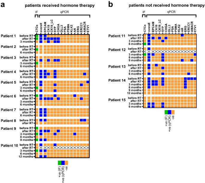
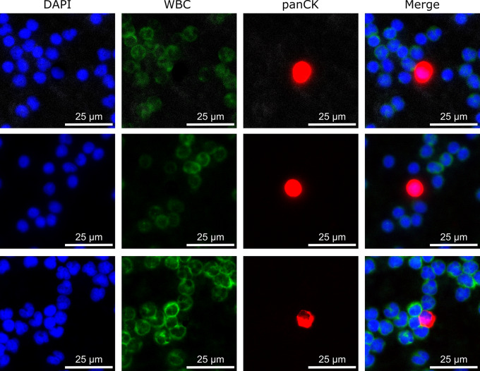
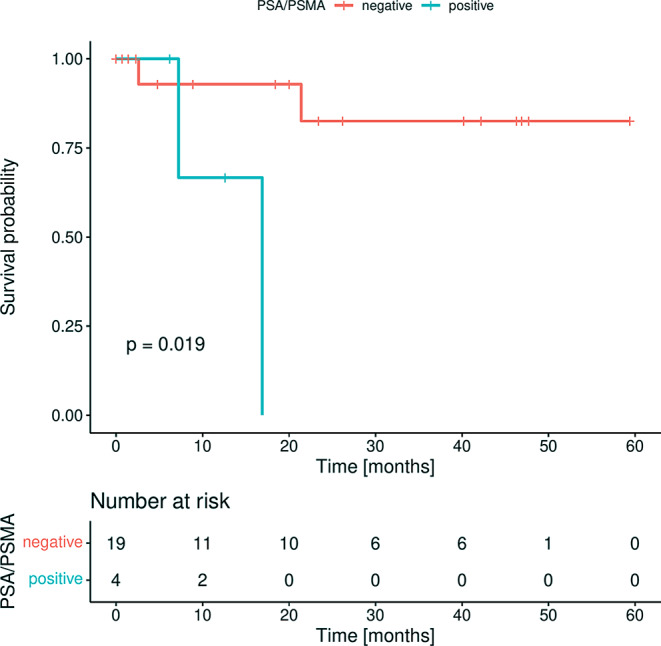
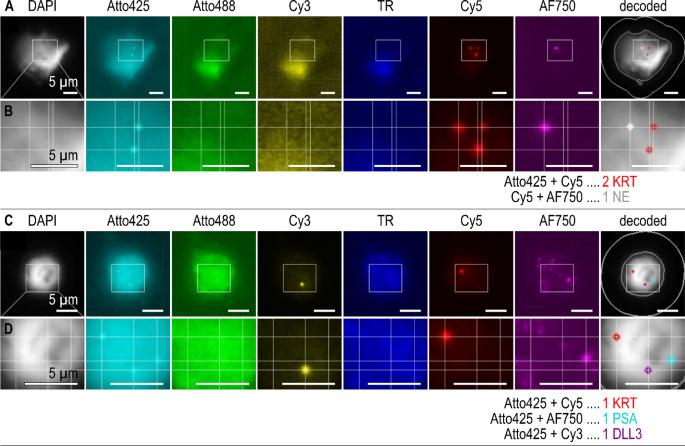
Circulating tumor cells (CTCs) are an established prognostic marker in metastatic prostate cancer (PrC) but have received little attention in localized high-risk disease. Peripheral blood was obtained from patients with early intermediate and high-risk PrC (n = 15) at baseline, after radiotherapy, and during follow-up, as well as from metastatic PrC patients (n = 23). CTCs were enriched using the microfluidic Parsortix® technology. CTC-related marker were quantified with qPCR and RNA in-situ hybridization (ISH). Positivity and associations to clinical parameters were assessed using McNemar test, Fisher Exact test or log-rank test. The overall positivity was high in both cohorts (87.0% metastatic vs. 66.7% early at baseline). A high concordance of qPCR and RNA ISH was achieved. In metastatic PrC, PSA and PSMA were prognostic for shorter overall survival. In early PrC patients, an increase of positive transcripts per blood sample was observed from before to after radiation therapy, while a decrease of positive markers was observed during follow-up. CTC analysis using the investigated qPCR marker panel serves as tool for achieving high detection rates of PrC patient samples even in localized disease. RNA ISH offers the advantage of confirming these markers at the single cell level. Employing the clinically relevant marker PSMA, our CTC approach can be used for diagnostic purposes to screen patients profiting from PSMA-directed PET-CT or PSMA-targeted therapy.

Keywords: Circulating tumor cells; Gene expression; Liquid biopsy; Parsortix; Prostate cancer; qPCR.


Conflict of interest statement

Declarations. Competing interest: The authors have no competing interest to declare.

Figures

Fig. 1
Fig. 1
Heatmap depicting positive and negative early PrC patients’ samples at time points before and after radiation therapy (RT), as well as after 3, 6, 9 or 12 months after treatment completion determined by qPCR CTC marker or CTC immunofluorescent (IF) staining. Patients that additionally received hormone therapy are shown at the left side (a) and patients that only received radiation therapy are shown on the right side (b) of the graph. qPCR positive samples (blue) were defined after applying a cut-off threshold. The detection of minimum one CTC was defined as detection limit for IF positive (green) samples. Negative samples by qPCR or IF are depicted in orange. Samples not available are marked with a cross (hydrolysis probe qPCR of patient 10 sample after radiotherapy could not be evaluated due to low expression of reference gene)
Fig. 2
Fig. 2
Example images of three different CTCs detected in early PrC patients by immunofluorescent staining of panCK (red), white blood cell (WBC) (green) and nuclear counterstaining using DAPI; A panCK positive, DAPI positive and WBC negative cell was defined as CTC; The given scale bar in each individual image marks the size of 25 μm
Fig. 3
Fig. 3
Heatmap depicting positive (blue) and negative (orange) metastatic PrC patient (n = 23) for qPCR CTC marker. qPCR positive samples were defined after applying a cut-off threshold
Fig. 4
Fig. 4
Kaplan-Meier plot for overall survival (OS) of metastatic prostate cancer patients according to PSA and PSMA positivity (blue) and negativity (red) in CTCs; Log-rank testing was used for comparing the patient’s outcome; p < 0.05 is defined as level of significance
Fig. 5
Fig. 5
Example images of two CTCs of metastatic PrC patient 2 detected by dual-color in-situ hybridization with (A-B) coexpression of cytokeratin (KRT) and neuroendocrine markers (NE (pool of CHGA, SYP, NCAM1)), and (C-D) coexpression of cytokeratin (KRT), prostate specific antigen (PSA) and neuroendocrine marker DLL3. In-situ signals were decoded based on their color code (e.g. colocalized Atto425 and Cy5 signals are decoded as KRT). Pseudocolored images are depicted for each channel, followed by a DAPI image with decoded in-situ signals and outlines of nucleus and cell border. Areas marked by white rectangles (A, C) are shown with higher magnification (B, D), grid lines for orientation help to identify and decode colocalized in-situ signals. The given scale bar in each individual image marks the size of 5 μm

References

    1. ECIS - European Cancer Information System European Union https://ecis.jrc.ec.europa.eu 20.7.2023
    1. American Cancer Society Cancer Statistics Center. http://cancerstatisticscenter.cancer.org/ 28.07.2023
    1. de Bono JS et al (2008) Circulating tumor cells predict survival benefit from treatment in metastatic castration-resistant prostate cancer. Clin Cancer Res 14 19:6302–6309 - PubMed
    1. Davis JW et al (2008) Circulating tumor cells in peripheral blood samples from patients with increased serum prostate specific antigen: initial results in early prostate cancer. J Urol 179 6:2187–2191 discussion 91 - PubMed
    1. Loh J et al (2014) Circulating tumor cell detection in high-risk non-metastatic prostate cancer. J Cancer Res Clin Oncol 140 12:2157–2162 - PMC - PubMed