Review
. 2024 Nov;44(11):1316-1336.
doi: 10.1002/cac2.12610. Epub 2024 Sep 21.

Altered glycosylation in cancer: molecular functions and therapeutic potential

Affiliations
Review

Altered glycosylation in cancer: molecular functions and therapeutic potential

Xuemeng Xu et al. Cancer Commun (Lond). 2024 Nov.

Abstract

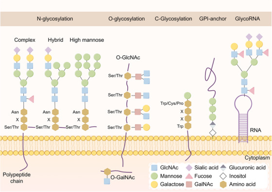
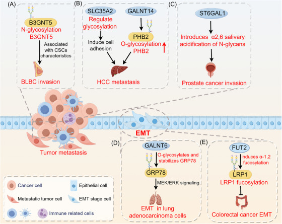
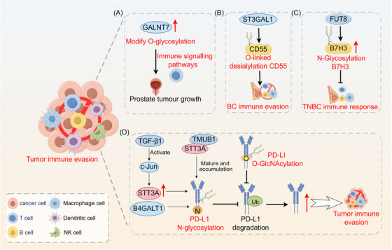
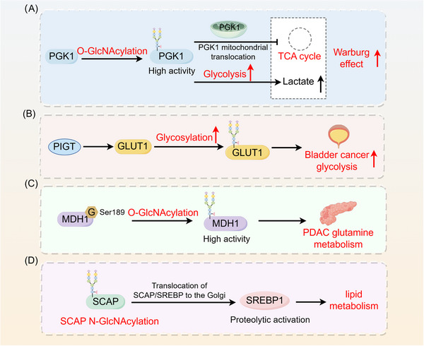
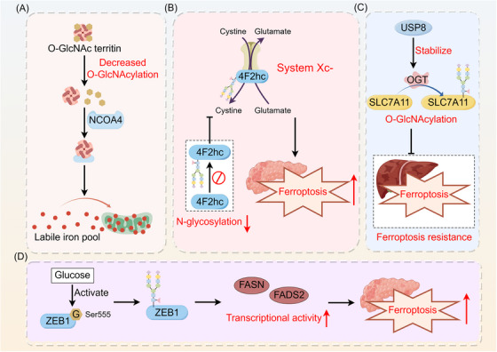
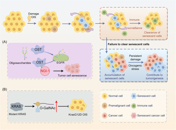
Glycosylation, a key mode of protein modification in living organisms, is critical in regulating various biological functions by influencing protein folding, transportation, and localization. Changes in glycosylation patterns are a significant feature of cancer, are associated with a range of pathological activities in cancer-related processes, and serve as critical biomarkers providing new targets for cancer diagnosis and treatment. Glycoproteins like human epidermal growth factor receptor 2 (HER2) for breast cancer, alpha-fetoprotein (AFP) for liver cancer, carcinoembryonic antigen (CEA) for colon cancer, and prostate-specific antigen (PSA) for prostate cancer are all tumor biomarkers approved for clinical use. Here, we introduce the diversity of glycosylation structures and newly discovered glycosylation substrate-glycosylated RNA (glycoRNA). This article focuses primarily on tumor metastasis, immune evasion, metabolic reprogramming, aberrant ferroptosis responses, and cellular senescence to illustrate the role of glycosylation in cancer. Additionally, we summarize the clinical applications of protein glycosylation in cancer diagnostics, treatment, and multidrug resistance. We envision a promising future for the clinical applications of protein glycosylation.

Keywords: Glycosylation; cancer therapy; cellular senescence; immunity; tumor biomarkers.


Conflict of interest statement

The authors declare that they have no competing interests.

Figures

FIGURE 1
FIGURE 1
Structural diversity of glycosylation. The types and numbers of oligosaccharide residues in the glycan chain, as well as the differences in the sites of glycosidic bonds, lead to the diversity of the glycan chains and glycosylations pathways. Depending on the glycosylation site, glycosylations are mainly classified as N‐glycosylation, O‐glycosylation, C‐glycosylation, or glycosylphosphatidylinositol anchoring. In addition to proteins and lipids, RNA is the third major carrier of glycosylation. Abbreviations: Asn, asparagine; Cys, cysteine; GalNAc, N‐acetylgalactosamine; GlcNAc, N‐acetylglucosamine; GlycoRNA, glycosylated RNA; GPI, glycosyl phosphatidylinositol; Ser, serine; Thr, threonine; Trp, tryptophan; X, any amino acid.
FIGURE 2
FIGURE 2
Role of glycosylation in tumor metastasis. Glycosylation has an impact on events related to tumor metastasis, including the stem cell properties of tumor cells, EMT, migration, and invasion. (A) B3GNT5 glycosylation is associated with cancer stem cell properties in basal‐like breast cancer. (B) SLC35A2 promotes HCC metastasis by regulating glycosylations to increase cell adhesion capacity. GALNT14‐mediated PHB2 O‐glycosylation promotes hepatocellular carcinoma cell growth and migration. (C) ST6GAL1 induces α2,6 salivary acidification of N‐glycans, promoting prostate cancer growth and invasion. (D) GALNT6 interacts with the O‐glycosylated chaperone protein GRP78, enhancing the MEK1/2/ERK1/2 signaling pathway in lung adenocarcinoma cells to promote EMT and invasion. (E) FUT2 induces α‐1,2 fucosylation and inhibits colorectal cancer EMT and metastasis via LRP1 fucosylation. Abbreviations: B3GNT5, β1,3‐N‐acetylglucosaminyltransferase 5; BLBC, basal‐like breast cancer; CSCs, cancer stem cells; EMT, epithelial‐mesenchymal transition; FUT2, fucosyltransferase 2; GALNT14, polypeptide N‐acetylgalactosaminyltransferase 14; GALNT6, N‐acetylgalactosaminyltransferase‐6; GRP78, glucose regulatory protein 78; HCC, hepatocellular carcinoma; LRP1, lipoprotein receptor‐related protein 1; PHB2, prohibitin 2; SLC35A2, solute carrier family 35 member A2.
FIGURE 3
FIGURE 3
Role of glycosylation in tumor immune evasion. (A) GALNT7 modifies O‐glycosylation, which is associated with the immune signaling pathways, in prostate cancer cells. (B) O‐linked salivary modification of CD55 by ST3GAL1 contributes to breast cancer cell immune evasion. (C) Abnormal B7H3 glycosylation mediated by FUT8 suppresses the immune response in TNBC cells. B4GALT1 mediates the N‐linked glycosylation of PD‐L1 protein, thereby preventing PD‐L1 degradation at the post‐transcriptional level. (D) TGF‐β1‐mediated glycosylation of PD‐L1 promotes immune evasion through the c‐Jun/STT3A signaling pathway. TMUB1 enhances PD‐L1 N‐glycosylation and stability by recruiting STT3A, thereby promoting PD‐L1 maturation and tumor immune evasion. O‐GlcNAcylation hinders the lysosomal degradation of PD‐L1 to promote tumor immune evasion. Abbreviations: B4GALT1, beta1,4‐galactosyltransferase 1; B7H3, B7 homolog 3 protein; BC, breast cancer; FUT8, fucosyltransferase 8; GALNT7, N‐acetylgalactosaminyltransferase 7; NK cell, natural killer cell; PD‐L1, programmed death‐ligand 1; ST3GAL1, ST3 beta‐galactoside alpha‐2,3‐sialyltransferase 1; STT3A, STT3 oligosaccharyl transferase complex catalytic subunit A; TGF‐β1, transforming growth factor‐beta1; TMUB1, transmembrane and ubiquitin‐like domain‐containing protein 1; TNBC, triple negative breast cancer; Ub, ubiquitin.
FIGURE 4
FIGURE 4
Role of glycosylation in tumor metabolic reprogramming. (A) Glycosylation increases the metabolic enzyme activity of PGK1 and induces PGK1 translocation to mitochondria to inhibit the TCA cycle, thereby enhancing the Warburg effect in cancer cells. (B) PIGT enhances glycolysis in bladder cancer cells through the regulation of GLUT1 glycosylation. (C) O‐GlcNAcylation regulates the metabolic activity of MDH1, promoting glutamine metabolism in pancreatic cancer. (D) SCAP N‐glycosylation promotes SCAP/SREBP translocation to the Golgi apparatus, which in turn activates SREBP1 to regulate lipid metabolism in tumors through SREBP‐dependent lipids. Abbreviations: G, glycosylation; GLUT1, glucose transporter type 1; MDH1, malate dehydrogenase 1; PDAC, pancreatic ductal adenocarcinoma; PGK1, phosphoglycerate kinase 1; PIGT, phosphatidylinositol glycan biosynthesis class T; SCAP, SREBP cleavage‐activating protein; SREBP, sterol regulatory element‐binding protein.
FIGURE 5
FIGURE 5
Role of glycosylation in tumor ferroptosis. Glycosylations interact with ferroptosis in cancer, modulating tumor progression. (A) Deglycosylation of the ferritin heavy chain enhances its interaction with the ferritin phagocytic receptor NCOA4, leading to the accumulation of unstable iron in mitochondria, which contributes to ferroptosis. (B) Inhibiting the N‐glycosylation of 4F2hc enhances the ferroptosis sensitivity of PDAC cells by suppressing the activity of the glutamate‐cystine reverse transport system Xc‐. (C) USP8 inhibits ferroptosis sensitivity in hepatocellular carcinoma by stabilizing OGT, which promotes the O‐GlcNAcylation of SLC7A11. (D) Glucose‐induced ZEB1 O‐GlcNAcylation activates the transcriptional activity of the adipogenesis‐related genes FASN and FADS2, leading to lipid peroxidation‐dependent ferroptosis in mesenchymal pancreatic cancer cells. Abbreviations: FADS2, fatty acid desaturase 2; FASN, fatty acid synthase; G, glycosylation; NCOA4, nuclear receptor coactivator 4; OGT, O‐GlcNAc transferase; PDAC, pancreatic ductal adenocarcinoma; SLC7A11, solute carrier family 7a member 11; USP18, ubiquitin‐specific peptidase 18; ZEB1, zinc finger E‐box binding homeobox 1.
FIGURE 6
FIGURE 6
Role of glycosylation in cellular senescence. Under certain conditions, the escape of senescent cells from immune surveillance may promote tumorigenesis and progression through mechanisms such as DNA damage and oxidative stress. (A) The cell‐permeable inhibitor NGI‐1 targets oligosaccharyl transferase and blocks cell surface localization and signaling of EGFR glycoproteins, promoting cellular senescence. (B) Increased levels of O‐GlcNAcylation in KRAS‐mutant lung cancer cells inhibit KrasG12D OIS and accelerate lung tumorigenesis. Abbreviations: EGFR, epidermal growth factor receptor; NGI‐1, N‐linked glycosylation inhibitor‐1; O‐GalNAc, O‐N acetylgalactosamine; OIS, oncogene‐induced senescence; OST, oligosaccharyl transferase.

References

    1. Wang WH, Yuan T, Qian MJ, Yan FJ, Yang L, He QJ, et al. Post‐translational modification of KRAS: potential targets for cancer therapy. Acta Pharmacol Sin. 2021;42(8):1201–1211. - PMC - PubMed
    1. Chen L, Liu S, Tao Y. Regulating tumor suppressor genes: post‐translational modifications. Signal Transduct Target Ther. 2020;5(1):90. - PMC - PubMed
    1. Gupta R, Sahu M, Srivastava D, Tiwari S, Ambasta RK, Kumar P. Post‐translational modifications: Regulators of neurodegenerative proteinopathies. Ageing Res Rev. 2021;68:101336. - PubMed
    1. Millan‐Zambrano G, Burton A, Bannister AJ, Schneider R. Histone post‐translational modifications ‐ cause and consequence of genome function. Nat Rev Genet. 2022;23(9):563–580. - PubMed
    1. Zhong Q, Xiao X, Qiu Y, Xu Z, Chen C, Chong B, et al. Protein posttranslational modifications in health and diseases: Functions, regulatory mechanisms, and therapeutic implications. Med Comm (2020). 2023;4(3):e261. - PMC - PubMed

Substances

Grants and funding