Review
. 2024 Oct 18;13(10):3069-3092.
doi: 10.1021/acssynbio.4c00219. Epub 2024 Sep 22.

Fundamentals and Exceptions of the LysR-type Transcriptional Regulators

Affiliations
Review

Fundamentals and Exceptions of the LysR-type Transcriptional Regulators

Wouter Demeester et al. ACS Synth Biol. .

Abstract

LysR-type transcriptional regulators (LTTRs) are emerging as a promising group of macromolecules for the field of biosensors. As the largest family of bacterial transcription factors, the LTTRs represent a vast and mostly untapped repertoire of sensor proteins. To fully harness these regulators for transcription factor-based biosensor development, it is crucial to understand their underlying mechanisms and functionalities. In the first part, this Review discusses the established model and features of LTTRs. As dual-function regulators, these inducible transcription factors exude precise control over their regulatory targets. In the second part of this Review, an overview is given of the exceptions to the "classic" LTTR model. While a general regulatory mechanism has helped elucidate the intricate regulation performed by LTTRs, it is essential to recognize the variations within the family. By combining this knowledge, characterization of new regulators can be done more efficiently and accurately, accelerating the expansion of transcriptional sensors for biosensor development. Unlocking the pool of LTTRs would significantly expand the currently limited range of detectable molecules and regulatory functions available for the implementation of novel synthetic genetic circuitry.

Keywords: LysR-type transcriptional regulators; biosensors; genetic circuitry; prokaryotes; synthetic biology; transcription factors.


Conflict of interest statement

The authors declare no competing financial interest.

Figures

Figure 1
Figure 1
(A) Schematic overview of transcription factor-based biosensors and their composition. For simplicity, the illustration is limited to inducible activators. The input is comprised of a transcription factor that can sense a ligand of interest. Signal processing occurs during DNA–protein interaction, where the transcription factor alters the expression of a target promoter depending on the input ligand concentration. This target promoter is responsible for the expression of an output gene that is chosen based on the envisioned biosensor application. (B, C) The two defining characteristics of these transcription factor-based biosensors are the response curve and ligand specificity. The response curve describes how biosensors link sensed ligand concentration to the output of choice. The ligand specificity determines how this response curve varies for different ligands. The response curves are given for a range of ligands, wherein the transcription factor exhibits low specificity (gray), as opposed to the high specificity observed for the ligand of interest (orange). Genetic circuit parts are given according to SBOL conventions., TF = transcription factor, TFBS = transcription factor binding site, FP = fluorescent protein, LOQ = limit of quantification, ULOQ = upper limit of quantification.
Figure 2
Figure 2
The regulatory mechanism of the LysR-type transcriptional regulators is referred to as the “sliding dimer” mechanism, owing to the movement of transcription factor dimers upon ligand interaction. (A) As most LysR-type transcriptional regulators (blue) are expressed from a bidirectional promoter region with one of their target genes (gray), this genetic architecture is given in more detail. (B) In the uninduced state, the tetrameric protein complex binds regulatory site (RS) and activation site 1 (AS1), causing the DNA to bend and preventing the RNA polymerase (RNAP) complex to recognize the promoter sequence. In addition, the location of the transcription factor binding sites overlap with key promoter regions (−10 and −35 boxes) of the target gene, further preventing RNAP complex–DNA interactions via steric hindrance. Due to overlap in the expression systems of the regulator and target gene, the transcription factor also causes negative autoregulation. Upon recognition of the appropriate ligand, the protein complex undergoes conformational changes that forces the dimer bound at AS1 to move to activation site 2 (AS2). These conformational changes are illustrated in a frontal and top view, showing the movement of the monomers within the tetrameric structure upon ligand interaction. As a result, the DNA bending is reduced and RNAP interaction sites become exposed, enabling activation of the target promoter transcription. On the surface of the ligand-bound regulator, RNAP recruitment sites appear after the conformational changes, further facilitating and guiding the interaction between RNAP and the target promoter. Genetic circuit parts are given according to SBOL conventions., TF = transcription factor, RS = regulatory site, AS = activation site, RNAP complex = RNA polymerase with appropriate sigma factor, LTTR = LysR-type transcriptional regulator.
Figure 3
Figure 3
Overview on the transcription factor binding sites of LysR-type transcriptional regulators. The target promoters of these regulators show three different binding sites, namely the regulatory site (RS) and two activation sites (AS1 and AS2). These regulatory sites follow the characteristic T-N11-A sequence, with symmetry in the nucleotides surrounding the thymine and adenine of this sequence. During DNA binding, the RS site is first bound by a dimer of regulator molecules. The high degree of symmetry in this site, as shown in bold for regulators BenM, DntR, and OccR, allows for a stable and strong interaction between the dimer and the DNA. A second dimer binds AS1, which shows a much lower degree of dyad symmetry. Hence, binding at this site is only possible while simultaneously forming the full tetrameric structure together with the RS-bound dimer. Due to the distance between RS and AS1, the tetramerization and DNA binding is accompanied by DNA bending. Similarly to the variation in AS1 sequence, AS2 also shows imperfect symmetry. The AS1 and AS2 sites cover important promoter regions, such as the −10 and −35 box. The location of these boxes, as well as the transcription start site, are given per promoter for both the target promoter system (gray) and the transcription factor expression system (blue). Genetic circuit parts are given according to SBOL conventions., TF = transcription factor, RS = regulatory site, AS = activation site.
Figure 4
Figure 4
(A) Annotation of the LysR-type transcriptional regulator (LTTR) protein. Using sequence and structural alignments, an amino acid consensus sequence was created, and specific boundaries of the protein domains were set. The sequence alignment was based on 134 unique and curated sequences from the Uniprot database (https://www.uniprot.org/), which can be found in Supplementary Table 1. The protein models used for the structural alignment were extracted from either the RCSB database (https://www.rcsb.org/) or the AlphaFold database (https://alphafold.com/), and are given in Supplementary Figure 1. (B) The structure of regulator CbnR is given, as well as a schematic illustration of the secondary structure of LTTRs, both annotated according to the domain definitions. The α-helices are given as squares and numbered from N-terminus to C-terminus, while the β-sheets are similarly given as arrows and letters A–F. Figure adapted from ref (265). DBD = DNA-binding domain, LH = linker helix, H = hinge, LBD = ligand-binding domain, RD = subdomain of the LBD, h = hydrophobic amino acids, X = any amino acid.
Figure 5
Figure 5
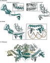
Oligomerization of CbnR as a model transcription factor for the LysR-type transcriptional regulators. (A) The monomers form two distinct conformations upon dimerization, i.e., the extended (left) and compact (right) form. These structural differences are stabilized by interacting residues between the linker helix and ligand-binding domain. A close-up of this region is given above the structures. (B) Dimerization occurs primarily around the linker helix in an antiparallel manner. As a result, the DNA-binding domains (DBDs) are positioned to enter two adjacent major grooves of the DNA, as shown in the box containing a close-up of the DBDs of CbnR in association with DNA. The wings of the winged helix-turn-helix structures interact with the nascent minor grooves. (C) Two dimers interact, forming the “dimer of dimers” conformations. The tetramerization interface is mainly located in the ligand-binding domains.
Figure 6
Figure 6
Overview of the currently identified alterations in LysR-type transcriptional regulators. (A) The components of the “classic” LysR-type transcriptional regulator model, given with “*”; their exceptions are summarized in five categories. Red arrows indicate repression, green arrows indicate activation, and gray arrows indicate no effect on transcription. (B) The possible effects of the exceptions described in A on the response curves of these regulators are visualized per parameter (as described in Figure 1B). Genetic circuit parts are given according to SBOL conventions.,T = Temperature, TF = transcription factor, TFBS = transcription factor binding site.

References

    1. Gardner T. S.; Cantor C. R.; Collins J. J. Construction of a genetic toggle switch in Escherichia coli. Nature 2000, 403, 339–342. 10.1038/35002131. - DOI - PubMed
    1. Elowitz M. B.; Leibler S. A synthetic oscillatory networkof transcriptional regulators. Nature 2000, 403, 335–338. 10.1038/35002125. - DOI - PubMed
    1. Cameron D. E.; Bashor C. J.; Collins J. J. A brief history of synthetic biology. Nature Reviews Microbiology 2014, 12, 381–390. 10.1038/nrmicro3239. - DOI - PubMed
    1. Zhang H.; Jiang T. Synthetic circuits, devices and modules. Protein and Cell 2010, 1, 974–978. 10.1007/s13238-010-0133-8. - DOI - PMC - PubMed
    1. Engstrom M. D.; Pfleger B. F. Transcription control engineering and applications in synthetic biology. Synthetic Syst. Biotechnol. 2017, 2, 176–191. 10.1016/j.synbio.2017.09.003. - DOI - PMC - PubMed

Publication types

MeSH terms

LinkOut - more resources