. 2024 Nov 1;202(5):734-744.
doi: 10.1667/RADE-24-00004.1.

56Fe-ion Exposure Increases the Incidence of Lung and Brain Tumors at a Similar Rate in Male and Female Mice

Affiliations

56Fe-ion Exposure Increases the Incidence of Lung and Brain Tumors at a Similar Rate in Male and Female Mice

Sophie R Finkelstein et al. Radiat Res. .

Abstract

The main deterrent to long-term space travel is the risk of Radiation Exposure Induced Death (REID). The National Aeronautics and Space Administration (NASA) has adopted Permissible Exposure Levels (PELs) to limit the probability of REID to 3% for the risk of death due to radiation-induced carcinogenesis. The most significant contributor to current REID estimates for astronauts is the risk of lung cancer. Recently updated lung cancer estimates from Japan's atomic bomb survivors showed that the excess relative risk of lung cancer by age 70 is roughly fourfold higher in females compared to males. However, whether sex differences may impact the risk of lung cancer due to exposure to high charge and energy (HZE) radiation is not well studied. Thus, to evaluate the impact of sex differences on the risk of solid cancer development after HZE radiation exposure, we irradiated Rbfl/fl, Trp53fl/+ male and female mice infected with Adeno-Cre with various doses of 320 kVp X rays or 600 MeV/n 56Fe ions and monitored them for any radiation-induced malignancies. We conducted complete necropsy and histopathology of major organs on 183 male and 157 female mice after following them for 350 days postirradiation. We observed that lung adenomas/carcinomas and esthesioneuroblastomas (ENBs) were the most common primary malignancies in mice exposed to X rays and 56Fe ions, respectively. In addition, 1 Gy 56Fe-ion exposure compared to X-ray exposure led to a significantly increased incidence of lung adenomas/carcinomas (P = 0.02) and ENBs (P < 0.0001) in mice. However, we did not find a significantly higher incidence of any solid malignancies in female mice as compared to male mice, regardless of radiation quality. Furthermore, gene expression analysis of ENBs suggested a distinct gene expression pattern with similar hallmark pathways altered, such as MYC targets and MTORC1 signaling, in ENBs induced by X rays and 56Fe ions. Thus, our data revealed that 56Fe-ion exposure significantly accelerated the development of lung adenomas/carcinomas and ENBs compared to X rays, but the rate of solid malignancies was similar between male and female mice, regardless of radiation quality.


Figures

FIG. 1.
FIG. 1.
Incidence of tumorigenesis after whole-body exposure to X rays vs. 56Fe ions in Rbfl/fl, Trp53fl/+ mice. Schematic representation of experimental design highlighting timeline for intranasal injection of Adeno-Cre virus, radiation exposure, and follow-up (panel A). Kaplan-Meier plots show malignancy-free survival (panel B) and multiple primary malignancies (MPM) or metastasis-free survival after whole-body exposure to 0, 1, 2, or 4 Gy of X rays (panel C). Pie graph shows the percentage incidence of adenoma/carcinoma, esthesioneuroblastoma, lymphoma, and other malignancies after whole-body X-ray exposure (panel D). Kaplan-Meier plots show malignancy-free survival (panel E) and multiple primary malignancies (MPM) or metastasis-free survival (panel F) after 0, 0.2, 0.5, or 1 Gy 56Fe-ions whole-body exposure. Pie graph shows the percentage incidence of adenoma/carcinoma, esthesioneuroblastoma, lymphoma, and other malignancies after whole-body 56Fe-ion exposure (panel G). n = number of mice. P values in Kaplan-Meier plots were calculated using Log-rank (Mantel-Cox) tests. *P < 0.05, **P < 0.01, ***P < 0.001, and ****P < 0.0001.
FIG. 2.
FIG. 2.
Incidence of tumorigenesis among male and female mice from second cohorts of mice. Bar graphs show the percentage incidence of any malignancies, lung adenoma/carcinoma and esthesioneuroblastoma after 0, 1, 2, or 4 Gy X-ray whole-body exposure. Bar graphs show the percentage incidence of any malignancies, lung adenoma/carcinoma, and esthesioneuroblastoma after 0, 0.2, 0.5, or 1 Gy 56Fe-ion whole-body exposure. Error bars represent the standard error of the mean calculated assuming a binomial distribution. P values in all bar graphs were calculated using chi-square tests by comparing the incidence of malignancies between males and females at each radiation dose. *P < 0.05, **P < 0.01, ***P < 0.001, and ****P < 0.0001.
FIG. 3.
FIG. 3.
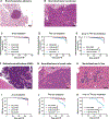
Incidence of bronchoalveolar adenoma/carcinoma and esthesioneuroblastoma (ENB) in unirradiated and irradiated cohorts. Panel A shows discrete, well-circumscribed bronchoalveolar adenoma (4×, bar = 200 μm). Panel B shows poorly circumscribed and infiltrative bronchoalveolar carcinoma is characterized by irregular lobules and clusters of neoplastic cells invading adjacent pulmonary tissue (4×, bar = 200 μm). Kaplan-Meier plots show bronchoalveolar adenoma/carcinoma-free survival after 0, 1, 2, or 4 Gy X-ray whole-body exposure (panel C), after 0, 0.2, 0.5, or 1 Gy of 56Fe-ion exposure (panel D), and 1 Gy X-ray exposure vs. 1 Gy 56Fe-ion exposure (panel E). Panel F shows large, invasive esthesioneuroblastoma effacing the olfactory bulb of the brain (4×, bar = 200 μm). Panel G shows olfactory neuroblastoma metastasis effacing the submandibular lymph node and compressing adjacent parotid (P) and sublingual (SL) salivary glands (4×, bar = 200 μm). Panel H shows metastatic clusters of olfactory neuroblastoma within liver sections (10×, bar =100 μm). Kaplan-Meier plots show the incidence of ENB-free survival after, 1, 2, or 4 Gy of X-ray whole-body exposure (panel I), 0, 0.2, 0.5, or 1 Gy of 56Fe ions (panel J), and 1 Gy of X rays vs. 1 Gy of 56Fe ions (panel K). n = number of mice. P values in Kaplan-Meier plots were calculated using Log-rank (Mantel-Cox) tests. *P < 0.05, **P < 00.01, ***P < 0.001, and ****P < 0.0001.
FIG. 4.
FIG. 4.
Esthesioneuroblastoma (ENB) whole transcriptome analysis using nanoString’s GeoMx digital spatial profiling. Panel A: Representative H&E and immunofluorescence images of the same samples (serial sections) utilizing fluorescent antibodies against NeuN (neuronal marker), GFAP (Astrocyte marker), and Syto 83 (Nuclei/DNA marker). Panel B: Heatmap shows unsupervised clustering of differentially expressed genes in control, X-ray and 56Fe-ion cohorts of ENBs. Volcano plots show differentially expressed genes in control vs. X-ray-induced ENBs (panel C) or control vs. 56Fe-ion-induced ENBs (panel D). Venn diagrams show a number of down- and up-regulated genes in X-ray- and 56Fe-ion-induced ENBs compared to the control (panel E). Hallmark pathway analysis shows significantly altered molecular pathways in control vs. X-ray-induced ENBs (panel F) or control vs. 56Fe-ion-induced ENBs (panel G). In volcano plots, P values associated with differentially expressed genes were found using the Wilcoxon test.

Update of

References

    1. Cucinotta F, Durante M. Cancer risk from exposure to galactic cosmic rays: Implications for space exploration by human beings. Lancet Oncol 2006; 7(5):431–5. - PubMed
    1. Schimmerling W, Cucinotta FA, Wilson JW. Radiation risk and human space exploration. Adv Space Res 2003; 31(1):27–34. - PubMed
    1. Durante M, Cucinotta FA. Heavy ion carcinogenesis and human space exploration. Nat Rev Cancer 2008; 8(6):465–72. - PubMed
    1. Cucinotta FA, Schimmerling W, Wilson JW, Peterson LE, Badhwar GD, Saganti PB, et al. Space radiation cancer risks and uncertainties for Mars missions. Radiat Res 2001; 156(5 Pt 2):682–8. - PubMed
    1. Preston DL, Ron E, Tokuoka S, Funamoto S, Nishi N, Soda M, et al. Solid cancer incidence in atomic bomb survivors: 1958–1998. Radiat Res 2007; 168(1):1–64. - PubMed

Publication types

LinkOut - more resources