. 2024 Sep 23;9(1):247.
doi: 10.1038/s41392-024-01960-8.

Single-cell exome sequencing reveals polyclonal seeding and TRPS1 mutations in colon cancer metastasis

Affiliations

Single-cell exome sequencing reveals polyclonal seeding and TRPS1 mutations in colon cancer metastasis

Jianqiang Cai et al. Signal Transduct Target Ther. .

Abstract

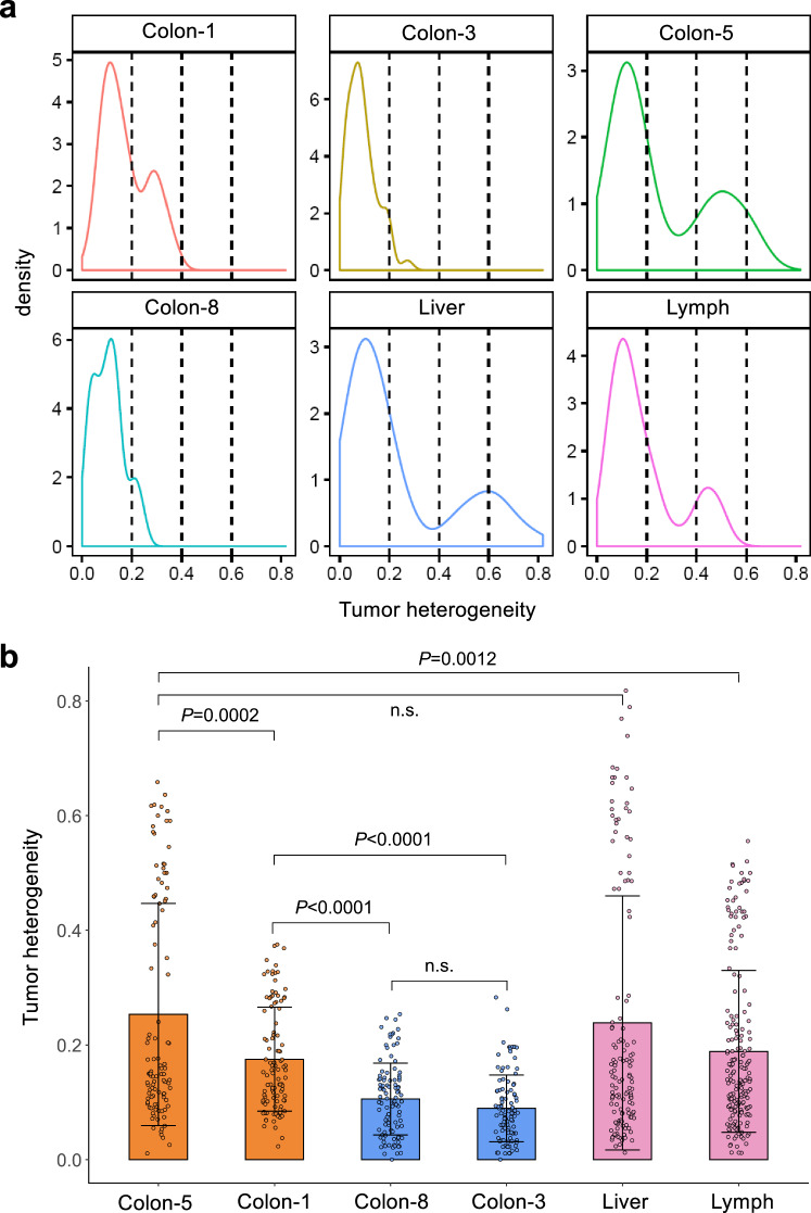
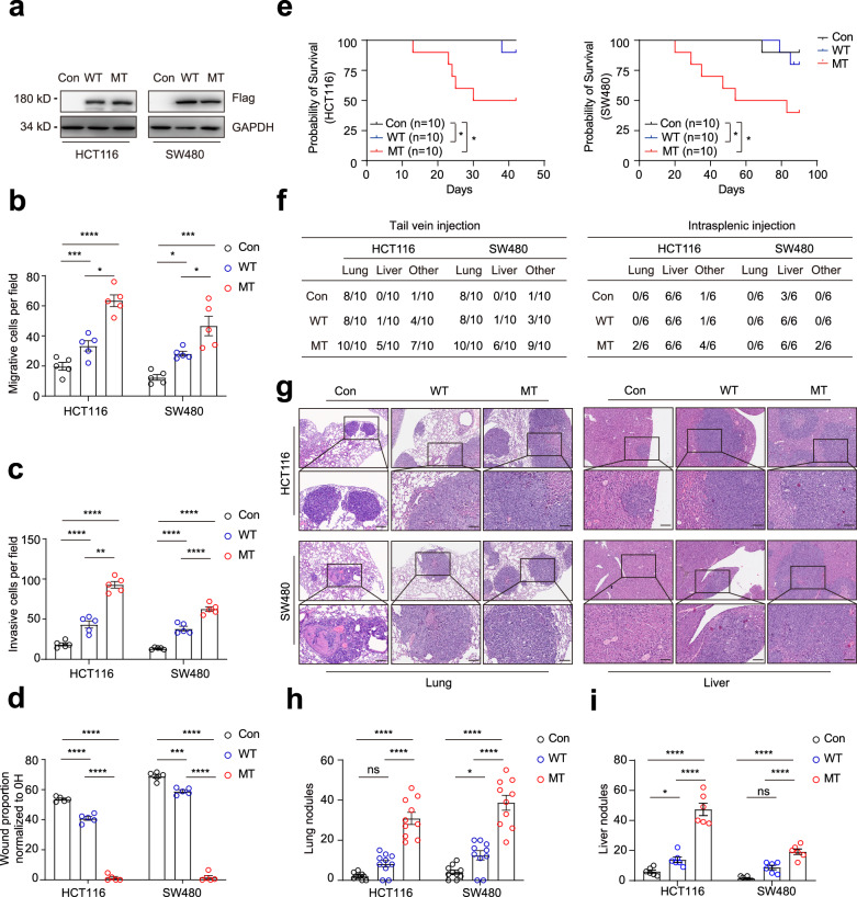
Liver metastasis remains the primary cause of mortality in patients with colon cancer. Identifying specific driver gene mutations that contribute to metastasis may offer viable therapeutic targets. To explore clonal evolution and genetic heterogeneity within the metastasis, we conducted single-cell exome sequencing on 150 single cells isolated from the primary tumor, liver metastasis, and lymphatic metastasis from a stage IV colon cancer patient. The genetic landscape of the tumor samples revealed that both lymphatic and liver metastases originated from the same region of the primary tumor. Notably, the liver metastasis was derived directly from the primary tumor, bypassing the lymph nodes. Comparative analysis of the sequencing data for individual cell pairs within different tumors demonstrated that the genetic heterogeneity of both liver and lymphatic metastases was also greater than that of the primary tumor. This finding indicates that liver and lymphatic metastases arose from clusters of circulating tumor cell (CTC) of a polyclonal origin, rather than from a single cell from the primary tumor. Single-cell transcriptome analysis suggested that higher EMT score and CNV scores were associated with more polyclonal metastasis. Additionally, a mutation in the TRPS1 (Transcriptional repressor GATA binding 1) gene, TRPS1 R544Q, was enriched in the single cells from the liver metastasis. The mutation significantly increased CRC invasion and migration both in vitro and in vivo through the TRPS1R544Q/ZEB1 axis. Further TRPS1 mutations were detected in additional colon cancer cases, correlating with advanced-stage disease and inferior prognosis. These results reveal polyclonal seeding and TRPS1 mutation as potential mechanisms driving the development of liver metastases in colon cancer.


Conflict of interest statement

The authors declare no competing interests.

Figures

Fig. 1
Fig. 1
Clustering and evolution analysis of adenoma, primary colon cancer, and metastatic tissues from a stage IV colon cancer patient. Single cells in the liver and lymph metastases could be tracked from colon 5 or colon 1. a Clustering of exome sequencing data of 140 single cells from primary tumor, liver metastasis, lymphatic metastasis, and adenoma from a stage IV colon cancer patient. Colon-N, normal tissue from colon; Colon-I, inner circle colon cancer cells; Colon-O, outer circle colon cancer cells. The height of the vertical lines in the dendrogram of graphs reflects the level at which two clusters are merged (based on the similarity of single cell). The greater the height, the more dissimilar the two clusters were at the point of their merger. b Heat-map of the 7 groups of genetic alterations with different distributions among the tissues. A red block in the “Damage” column indicates an SNV predicted to damage protein function. A purple block indicates a tolerated SNV. c Phylogenetic tree reconstruction depicting the evolutionary trajectory of colon cancer. The size of the nodes reflects the SNV frequency at each evolutionary stage, branches width reflects the frequency of SNV gain or loss, and branches reflects the number of SNV changes at each stage. Gene mutations marked in red are also found in the liver and lymph metastases. d A Sankey diagram shows the possible origins of liver and lymph metastasis based on single-cell exome sequencing of different tissue sections from the primary colon tumor (colon-1, 3, 5, 8). e The proportion of cells supporting the possible sites of origin of liver and lymph metastasis
Fig. 2
Fig. 2
Intra-tissue heterogeneity of the primary and metastatic tissues based on single-cell mutation landscape. a A density curve showing the distribution of intra-tissue heterogeneity within the primary and metastatic tissues. b A bar plot showing the difference in heterogeneity between the primary and metastatic tissues. Unpaired t-test, two-sided
Fig. 3
Fig. 3
Subclonal structure within 10 metastatic colon cancers and single-cell RNA-seq analysis of primary CRC and matched liver metastases samples of 6 CRLM patients. a Monoclonal seeding of 4 patients is shown as phylogenetic trees. b Polyclonal seeding of 4 patients is shown as phylogenetic trees. C: colon cancer (primary tumor), L: liver metastases, C + L: colon cancer and liver metastases. The relative branch length of the phylogenetic tree reflects the percentage of all mutations in a cluster. c UMAP plot segregating cancer cells of primary CRC into 8 subgroups. Colors indicate different subgroups. df UMAP plot showing the MC score, EMT score, CNV score of each single cell. Cells are colored according to MC score. g Sankey plots showing the associations between subgroups of cancer cells in primary CRC and matched liver metastases. h Histogram showing the proportion of tumor cells in primary CRC that metastasized to the liver in each CRLM patient. i Scatter plot with trend line showing a significant positive linear correlation between the EMT score and MC score
Fig. 4
Fig. 4
Intra-tissue heterogeneity of the primary and metastatic tissues based on the mutation status of APC. a The allele frequency of the APC stopgain mutation in the single cells from normal tissue, adenoma, colon-1, colon-3, colon-5, colon-8, liver metastasis and lymphatic metastasis. b The distribution of single cells categorized by homozygous mutant, heterozygous mutant, and wild-type APC. c The fractions of the APC mutation calculated from single cells correlate directly with mutation status based on the bulk tissue sequencing
Fig. 5
Fig. 5
TRPS1 R544Q mutation promotes colorectal cancer cells metastases in vitro and in vivo. a Western blot analyses of ectopic expression of TRPS1 and its mutant in HCT116 and SW480 cells. GAPDH serves as a loading control. (b, c) Graphs showing numbers of migrating or invasive cells expressing TRPS1 in HCT116 and SW480 cells using b transwell migration and c Matrigel invasion assays. d Bar graphs showing proportion of wound healing assay after 36 hours in the indicated HCT116 and SW480 cells. e Kaplan-Meier survival curves for BALB/c nude mice and NSG mice injected via tail vein with indicated HCT116 (n = 10) and SW480 cells (n = 10). f Table showing the incidence of metastasis in the lung, liver and other organs in mice. Other, additional organ metastases. g Representative images stained with H&E of lung (left) from tail vein models and liver (right) section from splenic intravenous injection models, featuring HCT116 (top) and SW480 (bottom) cells. Scale bars, 100 µm. Quantification of the number of h lung and i liver colonizing foci from the different groups. Data are represented as the mean ± SEM. Empty vector: Con, TRPS1 wild type: WT, TRPS1 mutant (R544Q): MT. All data are represented as the mean ± SEM, ns, not significant, *P < 0.05, **P < 0.01, ***P < 0.001, ****P < 0.0001
Fig. 6
Fig. 6
TRPS1 mutant (R544Q) promotes the colorectal cancer cell metastases through ZEB1. a Heatmap showing 172 common DEGs between HCT116 WT versus HCT116 MT (left) and SW480 WT versus SW480 MT (right) cells. Red and blue indicate high and low expression of genes. b The histogram of significantly enriched biological processes represented by the 172 DEGs from a generated through metascape functional enrichment analysis. c qRT-PCR and d western blot analysis showing mRNA and protein levels of ZEB1, SPARC, CDH1, and EpCAM in the indicated HCT116 and SW480 cells. e ChIP-qPCR analysis of TRPS1 enrichment at the ZEB1 promoter region in cells expressing TRPS1 WT and MT (R544Q). f Dual-luciferase reporter assay for co-transfection of TRPS1 WT/MT expression plasmids with ZEB1 promoter luciferase reporter constructs in 293 T cells. ZEB1 promoter fragments include the region from -2079/+200 and three truncated fragments (truncated fragment 1 [-2079~−1079], truncated fragment 2 [−1080~+200], truncated fragment 3 [−200~+200]. Bar graphs showing the number of migrating and invasive cells for HCT116 and SW480 cells with TRPS1 mutant/control and ZEB1 knockdown in g transwell migration and h Matrigel invasion assays. i Bar graph representing proportion of wound healing at 36 hours in HCT116 and SW480 cells with TRPS1 mutant/control and ZEB1 knockdown. j Representative images of immunohistochemistry for ZEB1 (upwards) and CDH1 (below) in CRC samples with TRPS1 wild type and mutations. Scale bars, 100 µm. k Quantification of ZEB1 (upwards) and CDH1 (below) H-scores in CRC samples with TRPS1 WT and all TRPS1 mutations. l Kaplan–Meier survival curves showing the correlation between TRPS1 mutant and DFS (upwards) or OS (below) in 107 CRC samples. Data are represented as the mean ± SEM. Empty vector: Con; TRPS1 wild type: WT; TRPS1 mutant (R544Q): MT. All data are represented as the mean ± SEM, ns, not significant, *P <0.05, ****P < 0.0001

References

    1. Mehlen, P. & Puisieux, A. Metastasis: a question of life or death. Nat. Rev. Cancer6, 449–458 (2006). - PubMed
    1. Hong, Y., Fang, F. & Zhang, Q. Circulating tumor cell clusters: What we know and what we expect (Review). Int. J. Oncol.49, 2206–2216 (2016). - PMC - PubMed
    1. Alexandrov, L. B. et al. Signatures of mutational processes in human cancer. Nature500, 415–421 (2013). - PMC - PubMed
    1. Kong, R. et al. Hypoxia-induced circ-CDYL-EEF1A2 transcriptional complex drives lung metastasis of cancer stem cells from hepatocellular carcinoma. Cancer Lett.578, 216442 (2023). - PubMed
    1. Naxerova, K. et al. Origins of lymphatic and distant metastases in human colorectal cancer. Science357, 55–60 (2017). - PMC - PubMed

Publication types