. 2024 Nov;11(43):e2403219.
doi: 10.1002/advs.202403219. Epub 2024 Sep 23.

A ROS-Responsive Lipid Nanoparticles Release Multifunctional Hydrogel Based on Microenvironment Regulation Promotes Infected Diabetic Wound Healing

Affiliations

A ROS-Responsive Lipid Nanoparticles Release Multifunctional Hydrogel Based on Microenvironment Regulation Promotes Infected Diabetic Wound Healing

Hao Yang et al. Adv Sci (Weinh). 2024 Nov.

Abstract

The continuous imbalance of the diabetic wound microenvironment is an important cause of chronic nonhealing, which manifests as a vicious cycle between excessive accumulation of reactive oxygen species (ROS) and abnormal healing. Regulating the microenvironment by suppressing wound inflammation, oxidative stress, and bacterial infection is a key challenge in treating diabetic wounds. In this study, ROS-responsive hydrogels are developed composed of silk fibroin methacrylated (SFMA), modified collagen type III (rCol3MA), and lipid nanoparticles (LNPs). The newly designed hydrogel system demonstrated stable physicochemical properties and excellent biocompatibility. Moreover, the release of antimicrobial peptide (AMP) and puerarin (PUE) demonstrated remarkable efficacy in eradicating bacteria, regulating inflammatory responses, and modulating vascular functions. This multifunctional hydrogel is a simple and efficient approach for the treatment of chronic diabetic infected wounds and holds tremendous potential for future clinical applications.

Keywords: antibacterial; antioxidation; anti‐inflammation; diabetic wound; multifunctional hydrogel.


Conflict of interest statement

The authors declare no conflict of interest.

Figures
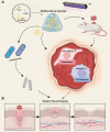
Figure 1
Figure 1
Diagrams displaying A) the formation of the SFMA/rColMA/LNP@AMP@PUE hydrogel and B) the mechanisms of accelerated wound healing.
Figure 2
Figure 2
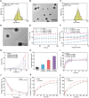
Characterization of SFMA/rColMA hydrogels loaded with ROS‐responsive LNPs. A) Particle size distribution of the LNPs. B) TEM images of the LNPs. Scale bar = 100 nm. C) Particle size distribution of LNP@AMP@PUE. D) TEM images of LNP@AMP@PUE. Scale bar = 100 nm. E) Rheological time‐scan curve of the hydrogel. F) Rheological frequency scanning curve of the hydrogel. G) Stress‒strain curves of the hydrogel. H) Compression module statistics under the maximum strain (n = 3). I) Swelling curve of the hydrogel (n = 3). J) Degradation curves of the 10% SFMA/2% rCol3MA/LNP@AMP@PUE hydrogel in PBS and enzyme environments (n = 3). K) Release rate of AMP in 10%SFMA/2%rCol3MA/LNP@AMP@PUE hydrogel. L) Release rate of PUE in 10%SFMA/2%rCol3MA/LNP@AMP@PUE hydrogel.
Figure 3
Figure 3
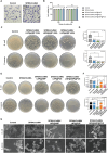
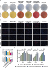
Evaluation of cytocompatibility and antibacterial capacity in vitro. A) The migration ability of fibroblasts in different groups was measured via a Transwell assay (n = 3). B) L929 cell viability after treatment with different hydrogels for 24 and 48 h (n = 3). C,D): Images and statistics of surviving bacterial clones after treatment with SFMA/rColMA hydrogels containing various concentrations of LNP@AMP (100, 200, or 400 µg mL−1) liposomes (n = 3). E), F): Images and statistics of surviving bacterial clones after treatment with different hydrogels (n = 3). G) SEM images of E. coli and S. aureus after different hydrogel treatments (white arrows indicate bacteria that ruptured after injury). Scale bar = 1 µm. ns indicates no significance, ** indicates p < 0.01, *** indicates p < 0.005.
Figure 4
Figure 4
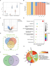
Analysis of the associations between diabetic wounds and PUE targets via network pharmacology. A) Distribution of the immune, stroma, and microenvironment scores of DFUs and normal skin. B) Fractions of macrophages and endothelial cells from DFUs and normal skin tissues. C) Volcano plot of DEGs from the GSE80178 dataset. D) Summary of target genes from different databases. E) Results of the KEGG pathway enrichment analysis results of target genes. F) Venn diagram of common genes. G) KEGG pathway enrichment analysis results of common genes. * indicates p < 0.05.
Figure 5
Figure 5
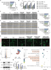
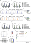
Effect of the SFMA/rColMA/LNP@AMP@PUE hydrogel on angiogenesis in the ROS microenvironment. A) Viability of HUVECs incubated with different hydrogels (n = 3). B) qRT‒PCR assay of the gene expression of angiogenesis‐related genes (CD31, Vcam1 and VWF) in HUVECs treated with different hydrogels (n = 3). C) Optical images of tube formation by HUVECs treated with different hydrogels. Scale bar = 100 µm. D), E): Quantification of the number of junctions and total length in each group (n = 3). F) Scratch assay images of HUVECs treated with different hydrogels. Scale bar = 100 µm. G) Quantification of the area change was performed (n = 3). H) Representative image and analysis of the mitochondrial membrane potential. JC‐1 was used to stain mitochondria with a strong membrane potential (red) and mitochondria with a weak membrane potential (green). Scale bar = 100 µm. I) Volcano map of the genes differentially expressed between the H2O2 group and the hydrogel‐treated group. J) Statistical plot of the differentially expressed genes. K) Pathway enrichment factor map of differentially expressed genes. L) Western blot analysis of HIF1‐α and VEGF expression in HUVECs after treatment with H2O2 and the SFMA/rColMA/LNP@AMP@PUE hydrogel. M) Quantitative analysis of HIF1‐α and VEGF expression via western blotting (n = 3). ** indicates p < 0.01, *** indicates p < 0.005, **** indicates p < 0.001.
Figure 6
Figure 6
Effect of the SFMA/rColMA/LNP@AMP@PUE hydrogel on the polarization of macrophages in the ROS microenvironment. A), B): qRT‒PCR analysis of the expression of typical M2 markers (IL‐10, ARG‐1, and CD206) and M1 markers (IL‐1β, iNOS, and CD80) in PMA‐pretreated THP‐1 cells treated with different hydrogels (n = 3). C), D), E), F): The flow cytometry results of PMA‐pretreated THP‐1 cells treated with different hydrogels were stained with the M1 marker CD86 and the M2 marker CD163 (n = 3). G,H): The expression levels of cytokines (TGF‐β, IL10, bFGF, IL‐1β, IL6, and TNF‐α) in PMA‐pretreated THP‐1 cells treated with NG/CMCS and NG/CMCS/HA/SF scaffolds were detected via ELISA (n = 3). I) Volcano map of the differentially expressed genes between the H2O2 group and the hydrogel‐treated group. J) Statistical plot of the differentially expressed genes. K) Pathway enrichment factor map of differentially expressed genes. L) Western blot analysis of AKT and p‐AKT expression in macrophages after treatment with H2O2 and the SFMA/rColMA/LNP@AMP@PUE hydrogel. M) Quantitative analysis of AKT and p‐AKT expression via western blotting (n = 3). ** indicates p < 0.01, *** indicates p < 0.005, **** indicates p < 0.001.
Figure 7
Figure 7
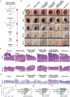
Assessment of wound healing in infected diabetic patients treated with different hydrogels. A) Treatment schedule for infected diabetic wounds treated with different hydrogels. B) Representative images of infected diabetic wounds at different times. C) HE staining of wound sections from all groups on Days 7 and 12. Scale bar = 1 mm. D) Masson staining images of wounds in all groups on Day 18. E) Quantification of the wound healing rate after treatment (n = 3). Scale bar = 1 mm. F) Statistics of granulation tissue thickness on Day 7 (n = 3). G) Collagen accumulation on Day 12, as determined via Masson's trichrome staining (n = 3). H) Statistical analysis of epidermal thickness on Day 18 (n = 3). * indicates p < 0.05, *** indicates p < 0.005, and **** indicates p < 0.001.
Figure 8
Figure 8
Evaluation of the antimicrobial ability and microenvironment regulation of the hydrogels in vivo. A,C): Images and statistics of surviving bacterial clones on Day 3 in vivo (n = 3). B), D): Immunofluorescence staining and statistical analysis of Ki67, Caspase 3, MPO, and CD68 expression in the wound tissues on Day 7 (n = 3). Scale bar = 50 µm. * indicates p < 0.05, *** indicates p < 0.005, and **** indicates p < 0.001.
Figure 9
Figure 9
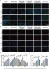
In vivo anti‐inflammatory and vascularization capacity of the hydrogels. A,C): Immunofluorescence images and statistics of IL4, TGFβ1, IFN‐γ, and TNF‐α in the wounds on Day 7 after different treatments (n = 3). Scale bar = 50 µm. B,D): Immunofluorescence images and statistics of CD31 and VEGF expression in the wounds on Day 7 after different treatments (n = 3). Scale bar = 50 µm. * indicates p < 0.05, *** indicates p < 0.005, and **** indicates p < 0.001.

References

    1. Howard B. V., Aragaki A. K., Tinker L. F., Allison M., Hingle M. D., Johnson K. C., Manson J. A. E., Shadyab A. H., Shikany J. M., Snetselaar L. G., Thomson C. A., Zaslavsky O., Prentice R. L., Diabetes Care 2018, 41, 680. - PMC - PubMed
    1. Weber M. B., Ranjani H., Staimez L. R., Anjana R. M., Ali M. K., Narayan K. M. V., Mohan V., Diabetes Care 2016, 39, 1760. - PMC - PubMed
    1. Guo J., Sun W., Kim J. P., Lu X., Li Q., Lin M., Mrowczynski O., Rizk E. B., Cheng J., Qian G., Acta Biomater. 2018, 72, 35. - PMC - PubMed
    1. Zhang J., Zhou R., Xiang C., Jia Q., Wu H., Yang H., Oxid. Med. Cell Longev. 2020, 2020, 4951820. - PMC - PubMed
    1. Singh V., Kashyap S., Yadav U., Srivastava A., Singh A. V., Singh R. K., Singh S. K., Saxena P. S., Toxicol. Res. (Camb) 2019, 8, 395. - PMC - PubMed

LinkOut - more resources