. 2024 May 24;13(9):103034.
doi: 10.1016/j.eats.2024.103034. eCollection 2024 Sep.

Modified Bridge-Enhanced Anterior Cruciate Ligament Repair

Affiliations

Modified Bridge-Enhanced Anterior Cruciate Ligament Repair

David E Kantrowitz et al. Arthrosc Tech. .

Abstract

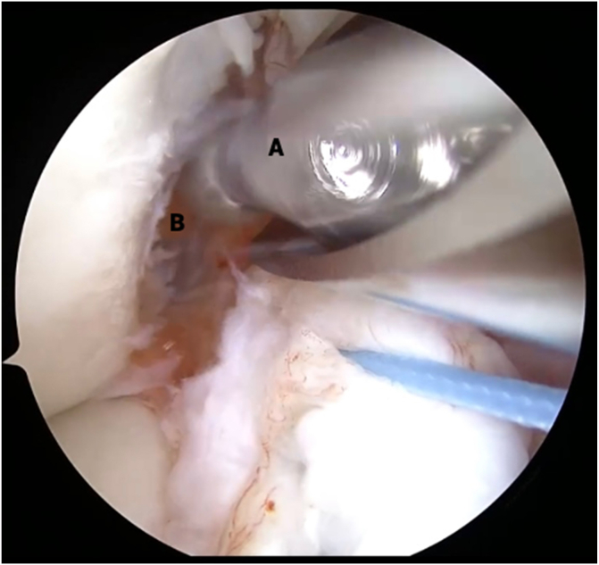
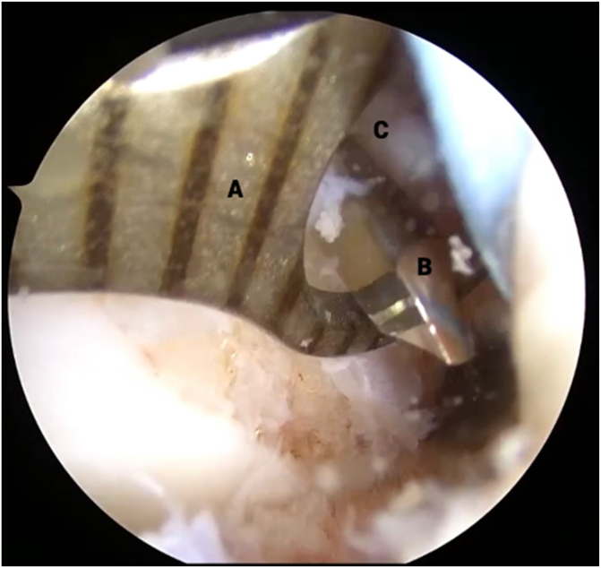
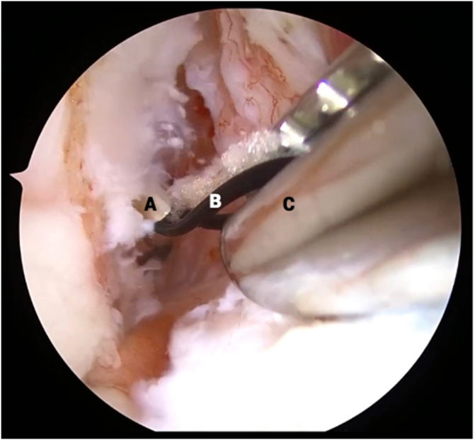
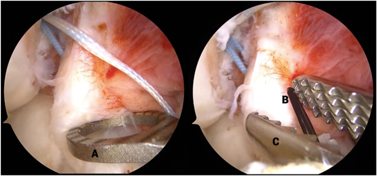
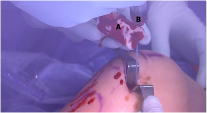
Historically, the treatment of anterior cruciate ligament (ACL) injuries shifted from primary repair to reconstruction because the native, intrasynovial location of the ACL precluded the formation of a fibrin-rich clot needed for ligament healing. However, increasing attention has been paid to augmenting the biological environment surrounding the ACL to facilitate its healing after arthroscopic repair. The bridge-enhanced ACL restoration implant uses resorbable collagen mixed with autologous blood to provide a biological scaffold for tissue healing. The short-term results of this procedure are promising, showing noninferiority to traditional ACL reconstruction at 2 years postoperatively and a higher rate of return to sport at 6 months. Our technique for performing the bridge-enhanced ACL repair is efficient, is easy to learn, and achieves excellent fixation of the ACL stump augmented with an internal brace.


Conflict of interest statement

The authors declare the following financial interests/personal relationships which may be considered as potential competing interests: J.N.G. reports a consulting or advisory relationship with DePuy Synthes Mitek Sports Medicine. S.G.A. reports a consulting or advisory relationship with Miach Orthopaedics. All other authors (D.E.K., C.N.D., E.D.H.) declare that they have no known competing financial interests or personal relationships that could have appeared to influence the work reported in this paper.

Figures

Fig 1
Fig 1
Preoperative setup. The patient is positioned supine on the operating table with a tourniquet on the right upper thigh (A), a foot bump to maintain the knee at 90° of flexion (C), and a lateral post to balance the knee when flexed (B).
Fig 2
Fig 2
View from anterolateral portal in right knee with supine positioning. An arthroscopic probe (B) is used to pull the torn anterior cruciate ligament (ACL) stump (C) away from the lateral intercondylar wall (A) to reveal the site of proximal avulsion. The torn ACL remnant tissue (C) is inspected for tear type, tissue height, vascularity, and tibial footprint integrity.
Fig 3
Fig 3
View from anterolateral portal in right knee with supine positioning. An arthroscopic tissue elevator (B) is used to carefully separate the anterior cruciate ligament (ACL) stump (A) from any scar tissue attachments. This step is critical to allow redirection of the proximal end of the ACL stump back to the native ACL footprint because scar attachments may prohibit superolateral translation of the stump tissue.
Fig 4
Fig 4
View from anterolateral portal in right knee with supine positioning. An arthroscopic suture passer (A) is used to suture the stump tissue (B) in an alternating, modified Bunnell configuration, beginning distally (shown on left) and working proximally (shown in middle), until both limbs of suture are exiting from the proximal end of the anterior cruciate ligament stump and the tissue is able to be reined with manipulation of the sutures (shown on right).
Fig 5
Fig 5
View from anterolateral portal in right knee with supine positioning. A 4.5-mm round burr (A) is used to roughen the lateral intercondylar ridge (B) to prepare for ligament implantation.
Fig 6
Fig 6
View from anteromedial portal in right knee with supine positioning. An outside-in drill guide (A) is brought in through the lateral portal and positioned over the femoral anterior cruciate ligament footprint. A small incision is made on the lateral thigh and deep through the iliotibial band so that a 2.7-mm drill (B) can be used to penetrate the lateral intercondylar notch (C) from outside in.
Fig 7
Fig 7
View from anterolateral portal in right knee with supine positioning. A looped nitinol wire (B) is passed through the drill sleeve (A), and an arthroscopic grasper (C) is used to retrieve the looped end of the wire out of the medial portal. The 2 anterior cruciate ligament repair sutures and 1 additional looped suture are then passed through the nitinol wire and shuttled out of the superolateral incision through which the femoral tunnel was made.
Fig 8
Fig 8
View from anterolateral portal in right knee with supine positioning. An outside-in drill guide (A) is positioned over the tibial footprint, and the tibia is penetrated with a 2.7-mm cannulated drill, through which a looped nitinol wire is passed (B) and retrieved with an arthroscopic grasper (C) out of the medial portal.
Fig 9
Fig 9
Clinical view of right knee, positioned supine, during internal brace application. The anterior cruciate ligament repair sutures (A) are loaded onto a titanium button (B) that is preloaded with a Minitape suture (C) functioning as an internal brace. The free limbs of the internal brace are passed through the looped passing suture (D), pulled into the superolateral incision (E), and shuttled out of the medial portal.
Fig 10
Fig 10
View from anterolateral portal in right knee with supine positioning. The anterior cruciate ligament (A) and internal brace (B) are seen in tensioned femorotibial orientation prior to bridge-enhanced anterior cruciate ligament restoration (BEAR) implantation.
Fig 11
Fig 11
Clinical view of right knee, positioned supine, during capsulotomy step. A No. 11 blade scalpel (A) is used to extend the medial portal (B) proximally until the notch can be clearly visualized to prepare for bridge-enhanced anterior cruciate ligament restoration (BEAR) insertion. Army-navy retractors (C) are used for careful dissection to avoid injury to the meniscus, medial femoral condyle, or internal brace sutures (D).
Fig 12
Fig 12
Clinical view of right knee, positioned supine, during bridge-enhanced anterior cruciate ligament restoration (BEAR) suture passage. Each limb of the internal brace is loaded onto a straight free needle (A) and individually passed through the center of the BEAR implant (B).
Fig 13
Fig 13
Clinical view of right knee, positioned supine, during hydration of bridge-enhanced anterior cruciate ligament restoration (BEAR) implant. Ten milliliters of the patient’s blood is collected by the anesthesiologist and handed off in a sterile syringe (B). The BEAR implant (A) is hydrated with the patient’s blood, beginning with 2 mL centrally, followed by the remaining 8 mL peripherally. The distal end of the implant is left dehydrated to allow for a stiffer surface against which to push the implant into the notch.
Fig 14
Fig 14
Clinical view of right knee, positioned supine, postoperatively after closure of incisions.

References

    1. Sanders T.L., Maradit Kremers H., Bryan A.J., et al. Incidence of anterior cruciate ligament tears and reconstruction: A 21-year population-based study. Am J Sports Med. 2016;44:1502–1507. - PubMed
    1. Herzog M.M., Marshall S.W., Lund J.L., Pate V., Mack C.D., Spang J.T. Trends in incidence of ACL reconstruction and concomitant procedures among commercially insured individuals in the United States, 2002-2014. Sports Health. 2018;10:523–531. - PMC - PubMed
    1. Feagin J.A., Curl W.W. Isolated tear of the anterior cruciate ligament: 5-Year follow-up study. Am J Sports Med. 1976;4:95–100. - PubMed
    1. Odensten M., Hamberg P., Nordin M., Lysholm J., Gillquist J. Surgical or conservative treatment of the acutely torn anterior cruciate ligament: A randomized study with short-term follow-up observations. Clin Orthop Relat Res. 1985;198:87–93. - PubMed
    1. Sandberg R., Balkfors B., Nilsson B., Westlin N. Operative versus non-operative treatment of recent injuries to the ligaments of the knee. A prospective randomized study. J Bone Joint Surg Am. 1987;69:1120–1126. - PubMed

LinkOut - more resources