

Neuroschistosomiasis: A Case Report and Review of Literature
- PMID: 39309391
- PMCID: PMC11412587
- DOI: 10.4103/jwas.jwas_174_23
Neuroschistosomiasis: A Case Report and Review of Literature
Abstract
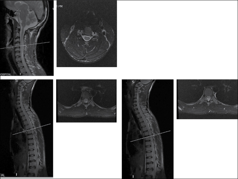
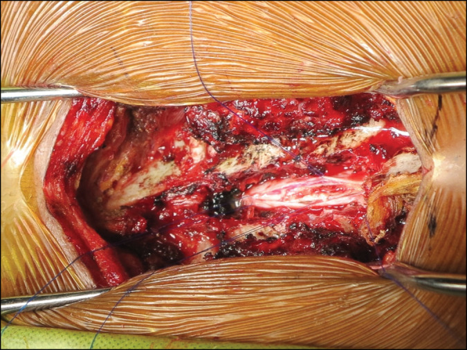
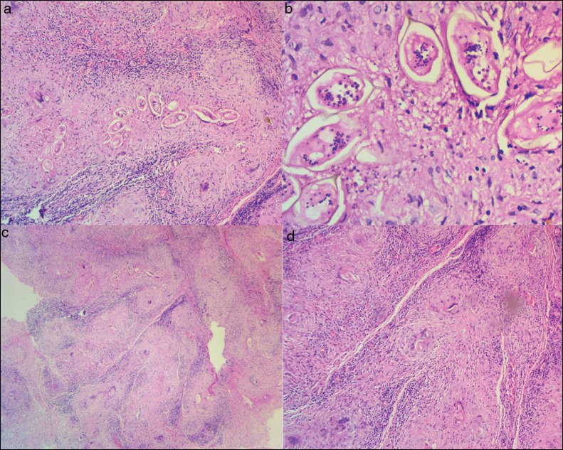
Spinal cord involvement is a rare complication of the schistosomiasis manifesting as myeloradiculopathy, medullary or conus-cauda equina syndrome which can lead to potentially serious long-term disability. Computed tomography and magnetic resonance imaging coupled with biochemical parameters have become the mainstay of diagnosis. Biopsy which is the gold standard of diagnosis demonstrating the organism is usually reserved for cases of diagnostic challenge. We report a rare case of upper thoracic spinal cord schistosomiasis diagnosed by biopsy in an 18-year-old male migrant presenting to a spine and orthopaedic centre in Ghana with complaints of upper back pain and associated myeloradiculopathy symptoms. Initial suspicion of intramedullary cord tumour was made based on magnetic resonance imaging findings warranting biopsy which revealed schistosoma spp. He was treated with anthelminthics and corticosteroids with a resolution of symptoms.
Keywords: Anthelminthics; back; corticosteroids; myeloradiculopathy; pain; schistosomiasis; spinal cord; thoracic.
Copyright: © 2024 Journal of the West African College of Surgeons.
Conflict of interest statement
The authors declare no conflicts of interest.
Figures
References
-
- Luyendijk W, Lindeman J. Schistosomiasis (bilharziasis) mansoni of the spinal cord stimulating an intramedullary tumor. Surg Neurol. 1975;4:457–60. - PubMed
-
- Dar J, Zimmerman RR. Schistosomiasis of the spinal cord. Surg Neurol. 1977;8:416–8. - PubMed
-
- Ferrari TCA, Moreira PRR. Neuroschistosomiasis: Clinical symptoms and pathogenesis. Lancet Neurol. 2011;10:853–64. - PubMed
-
- Vale TC, de Sousa-Pereira SR, Ribas JGR, Lambertucci JR. Neuroschistosomiasis mansoni: Literature review and guidelines. Neurologist. 2012;18:333–42. - PubMed
Publication types
LinkOut - more resources
Full Text Sources
Miscellaneous