. 2024 Sep;14(9):4028-4044.
doi: 10.1016/j.apsb.2024.05.026. Epub 2024 May 31.

Identification of novel small-molecule inhibitors of SARS-CoV-2 by chemical genetics

Affiliations

Identification of novel small-molecule inhibitors of SARS-CoV-2 by chemical genetics

Chris Chun-Yiu Chan et al. Acta Pharm Sin B. 2024 Sep.

Abstract

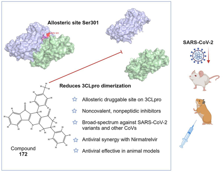
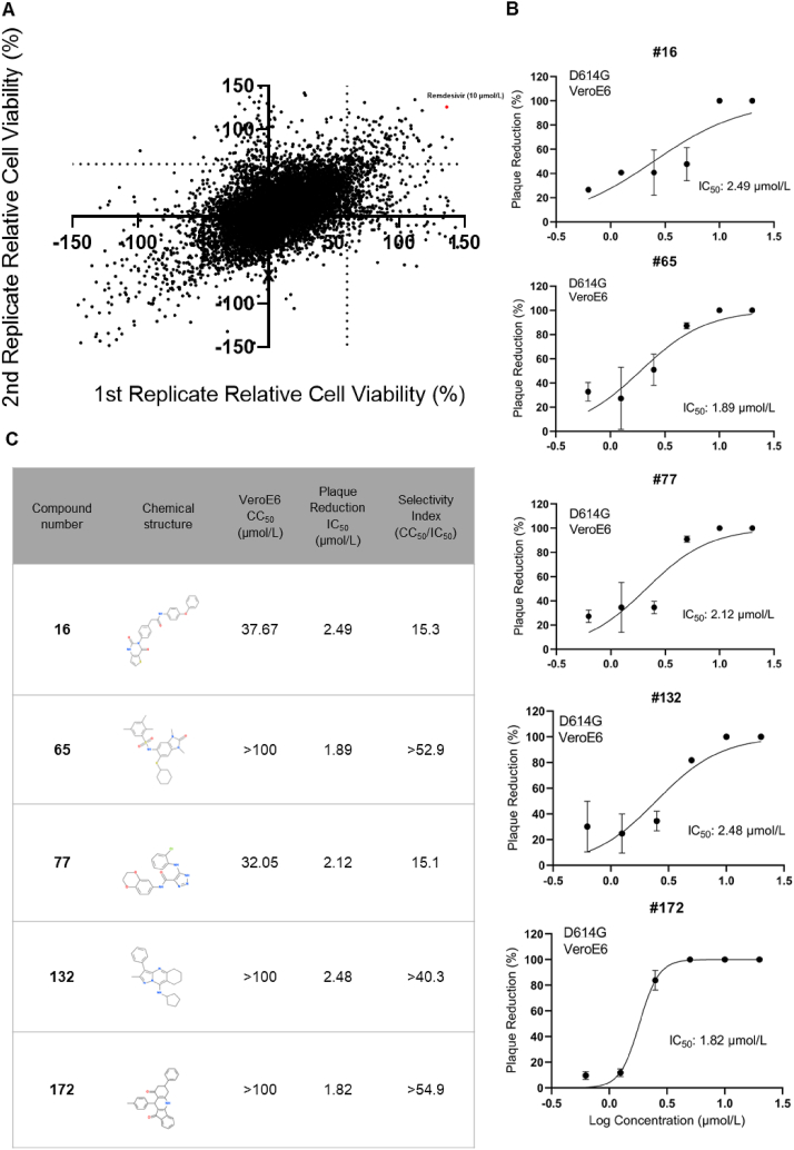
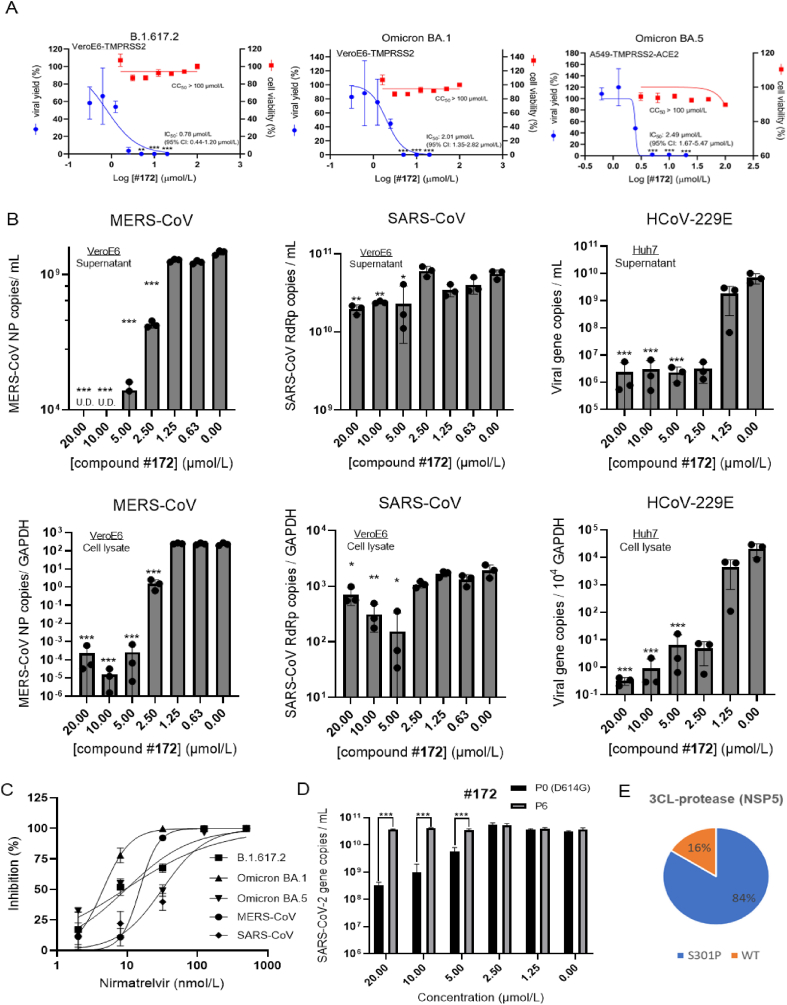
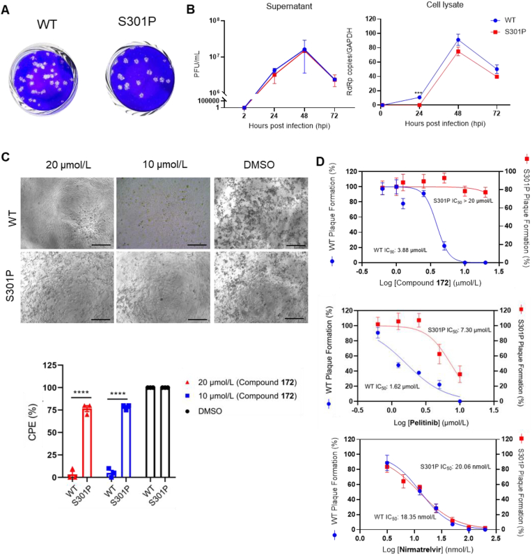
There are only eight approved small molecule antiviral drugs for treating COVID-19. Among them, four are nucleotide analogues (remdesivir, JT001, molnupiravir, and azvudine), while the other four are protease inhibitors (nirmatrelvir, ensitrelvir, leritrelvir, and simnotrelvir-ritonavir). Antiviral resistance, unfavourable drug‒drug interaction, and toxicity have been reported in previous studies. Thus there is a dearth of new treatment options for SARS-CoV-2. In this work, a three-tier cell-based screening was employed to identify novel compounds with anti-SARS-CoV-2 activity. One compound, designated 172, demonstrated broad-spectrum antiviral activity against multiple human pathogenic coronaviruses and different SARS-CoV-2 variants of concern. Mechanistic studies validated by reverse genetics showed that compound 172 inhibits the 3-chymotrypsin-like protease (3CLpro) by binding to an allosteric site and reduces 3CLpro dimerization. A drug synergistic checkerboard assay demonstrated that compound 172 can achieve drug synergy with nirmatrelvir in vitro. In vivo studies confirmed the antiviral activity of compound 172 in both Golden Syrian Hamsters and K18 humanized ACE2 mice. Overall, this study identified an alternative druggable site on the SARS-CoV-2 3CLpro, proposed a potential combination therapy with nirmatrelvir to reduce the risk of antiviral resistance and shed light on the development of allosteric protease inhibitors for treating a range of coronavirus diseases.

Keywords: 3CLpro inhibitor; Allosteric-site inhibitor; Animal models; Broad-spectrum antiviral treatment; Chemical genetics; High throughput screening; Reverse genetics; SARS-CoV-2.


Conflict of interest statement

The authors have no conflict to declare.

Figures

Image 1
Graphical abstract
Figure 1
Figure 1
Phenotype-based high-throughput antiviral screening. (A) Primary screening results using a CPE-inhibition assay. The primary screening cutoff was set as over 60% cell viability in both duplicates. Out of 50,213 compounds, 168 were identified to satisfy this criterion. Mock infection was included to normalize the cell viability (100%). Remdesivir was added at a concentration of 10 μmol/L as positive control (shown in red). (B) Plaque reduction assay results of the five finalized compounds. VeroE6 cells were inoculated with 40 PFU of SARS-CoV-2 D614G virus and subsequently treated with each of the five selected hit compounds, which were diluted from 20 to 1.25 μmol/L in 2-fold intervals. At 72 hpi, the cells were fixed with 10% formalin/PBS and stained with a 0.5% crystal violet solution to visualize the plaques. The IC50 value was calculated by fitting the number of plaque formations against the logarithmic concentrations of the compounds using a non-linear regression model in GraphPad Prism software. (C) Antiviral profiles of the five selected compounds with their chemical structure, IC50 (by plaque reduction assay), CC50 (by MTT cytotoxicity assay), and selectivity index (SI) as shown. The highest tested concentration in the MTT cytotoxicity assay was 100 μmol/L due to the maximal water solubility in 1% DMSO.
Figure 2
Figure 2
Pan-coronavirus activity and raise of escape mutant of compound 172. (A) viral load reduction assay conducted against SARS-CoV-2 VOC Delta, Omicron BA.1/BA.5, and co-plotted with cytotoxicity using MTT cytotoxicity assays. The viral yield was expressed as a percentage of DMSO control. Compound 172 was diluted in 2-fold intervals from 20 μmol/L to 0.3125 μmol/L, and the IC50 value was calculated by fitting the normalized viral load against the logarithmic concentrations using a non-linear regression model in GraphPad Prism software. MTT cytotoxicity assays were carried out as previously described. (B) VeroE6 or Huh7 cells were infected with MERS-CoV, SARS-CoV, or HCoV-229E at MOI 0.01. After 48 hpi, the supernatant and cell lysate were collected and lysed, and the viral RNA copies were measured using RT-qPCR. (C) Antiviral activity of Nirmatrelvir as a reference control of compound 172. (D) VeroE6-TMPRSS2 cells were infected either by Passage 0 (D614G virus) or Passage 6 virus at MOI 0.01, followed by treatment with varying concentrations of compound 172. After 48 h, the supernatant was collected and the viral load was measured using RT-qPCR. Statistical analysis for all the assays above were performed by Student's t-test: Data are presented as mean ± SD. ∗∗∗P < 0.001; ∗∗P < 0.01; ∗P < 0.05. (E) Nanopore sequencing results. 84% virus population carried S301P point mutation in the 3CLpro. 172 Escape mutant was raised in six passages in VeroE6-TMPRSS2, by reducing MOI and increasing 172 concentrations in each passage, until the CPE inhibition effect of 172 was abolished.
Figure 3
Figure 3
Characterization of compound 172-resistant S301P recombinant SARS-CoV-2. (A) Plaque morphology of recombinant WT and 3CLpro S301P virus. VeroE6-TMPRSS2 cells were utilized to culture and quantitate 3CLpro S301P virus, which was rescued using VeroE6-TMPRSS2 cells. (B) Replication kinetics of 3CLpro S301P recombinant virus. Both recombinant WT and mutant viruses replicate at comparable rate, 3CLpro S301P showed slight attenuation in growth rate. (C) Microscopic images of recombinant WT and 3CLpro S301P mutant virus in the presence of compound 172 and percentages of CPE of each image. VeroE6-TMPRSS2 was infected with either virus at MOI 0.01 and treated with compound 172 at different concentrations ranging from 20 to 0.625 μmol/L. Representative images of CPE formation were captured at 48 hpi. Scale bar = 100 μm. The experiment was performed in triplicate and repeated twice for confirmation. The CPE severity in each group was scored and compared by two-way ANOVA. (D) Plaque formation assay of recombinant WT and 3CLpro S301P virus under the treatment of compound 172, Pelitinib, and Nirmatrelvir, respectively. Antiviral IC50 of each drug compound against WT and mutant viruses were plotted by GraphPad. Data are presented as mean ± SD. ∗∗∗∗P < 0.0001; ∗∗∗P < 0.001.
Figure 4
Figure 4
Mechanistic investigation of compound 172. (A) FRET-based protease activity assay. Recombinant WT and S301P 3CLpro were expressed and purified. Compound 172 was diluted from 100 to 6.26 μmol/L and incubated with 1.25 μmol/L WT or S301P 3CLpro at RT for 30 min. 50 μmol/L fluorophore-conjugated substrate was added to the mixture after 20 min as previously established. 200 nmol/L GC376 was added as a positive control. Both experiments were performed in triplicate. Statistical analysis by Student's t-test: Data present as mean ± SD. ∗∗∗P < 0.001; ∗∗P < 0.01; ∗P < 0.05. (B, C) Surface plasmon resonance (SPR) spectroscopy of compound 172 with WT and S301P 3CLpro. The Biacore T-200 machine was used to conduct SPR spectroscopy. 40 μg/mL of protease was immobilized on a Series S Sensor Chip CM5. Compound 172 was added to the chip at gradient concentrations ranging from 100 to 3.125 μmol/L. Cytiva software was used to generate the association-dissociation graph and the dissociation constant (KD). (D, E) Michaelis–Menton inhibition kinetics of compound 172 and Nirmatrelvir. 1.25 μmol/L WT 3CLpro was incubated with compound 172 or Nirmatrelvir in a gradient of concentrations at RT for 30 min, followed by the addition of fluorophore-conjugated substrate in a range of concentrations and measured in fluorescence (Excitation: 365 nm, Emission: 500–550 nm) in 20 min as initial velocity. (F) Analytical ultracentrifugation (AUC) for WT 3CLpro with compound 172. 600 μg/mL WT 3CLpro was incubated with 20 μmol/L compound 172 or 0.2% DMSO before this sedimentation velocity experiment. The main peaks for 3CLpro monomer and dimer showed a sedimentation coefficient of 3.3 and 4.6 S respectively. Two independent experiments were performed. Statistical analysis by Student's t-test: Data are presented as mean ± SD. ∗∗∗P < 0.001; ∗∗P < 0.01; ∗P < 0.05. (G, H) Intermolecular interaction of pelitinib and compound 172 to 3CLpro. Monomers are shown in cyan and green cartoon representations, respectively. Interacting residues of 3CLpro within 3.5 Å of compound are shown in magenta sticks and labelled accordingly. (I) List of Loewe's additivity (LA) indices calculated with the IC50 values of each compound combination. LA < 1, synergism; LA = 1, independent; LA > 1, antagonism. 172 concentrations that lowered Nirmatrelvir IC50 are highlighted in green. This experiment was conducted in triplicates.
Figure 5
Figure 5
In vivo activity of compound 172. (A) Permeability of compound 172 in Caco-2 cells and metabolic stability of 172 in mouse and human Liver microsomes. (B) In vivo pharmacokinetic profiling of compound 172 in a BALB/c mouse model (n = 3). (C) Schematics of compound 172 administration in a golden Syrian hamster model. Two groups of hamsters (n = 4) were given intranasal inoculations of 1000 PFU/animal of WT SARS-CoV-2. The treatment group (n = 4) received compound 172 (1 mg/kg) or nirmatrelvir (200 mg/kg) via intraperitoneal (ip) injection, or pelitinib (10 mg/kg) via oral gavage. The vehicle group (n = 4) was given 5% DMSO in 20% SBE-β-CD/0.9% saline. On the third day following the infection, the hamsters were euthanized for both viral yield and histopathological examination. (D) Hamsters lung and nasal turbinate viral yield were determined by RT-qPCR. Statistical analysis by one-way ANOVA: Data present as mean ± SD. ∗∗∗P < 0.001; ∗∗P < 0.01; ∗P < 0.05. (E) Representative images of H&E-stained and IF-stained lung tissue sections from hamster treated as indicated. Scale bars: 100 μm. (F) Schematic of compound 172 administration in a K18-hACE2 mouse model. K18-hACE2 mice were intranasally inoculated with either Alpha (B.1.1.7, n = 5 for survival rate) or Omicron (BA.5, n = 4 for viral load detection) at 200 PFU/animal or 10,000 PFU/animal, respectively. The treatment group was given compound 172 (50 mg/kg) dissolved in a solution of 20% SBE-β-CD/0.9% saline, administered once daily via i.p. injection. The vehicle group was given 5% DMSO in 20% SBE-β-CD/0.9% saline using the same treatment regimen. The positive control group was given Nirmatrelvir (200 mg/kg) dissolved in a solution of 20% SBE-β-CD and 0.9% saline using the same treatment regimen. The mice in the survival study were monitored daily, and drugs were administered until they reached the humane endpoint or died. The mice in the viral load study were euthanized on the third day after the infection, and their lungs and nasal turbinate were collected for viral load quantification. (G) Mouse survival rate (upper panel) and daily body weight changes (lower panel) of K18-hACE2 mice. The comparison of survival rates between groups were analysed using Log-rank (Mantel–Cox) tests and that of body weight using two-way ANOVA. (H) K18-hACE2 mouse lung and nasal turbinate viral titer determined by standard plaque assay. Statistical analysis by one-way ANOVA: Data are presented as mean ± SD. ∗∗∗P < 0.001; ∗∗P < 0.01; ∗P < 0.05; ns indicates not significant.

Similar articles

Cited by

References

    1. Peiris J.S., Lai S.T., Poon L.L., Guan Y., Yam L.Y., Lim W., et al. Coronavirus as a possible cause of severe acute respiratory syndrome. Lancet. 2003;361:1319–1325. - PMC - PubMed
    1. Zaki A.M., van Boheemen S., Bestebroer T.M., Osterhaus A.D., Fouchier R.A. Isolation of a novel coronavirus from a man with pneumonia in Saudi Arabia. N Engl J Med. 2012;367:1814–1820. - PubMed
    1. Zhou P., Yang X.L., Wang X.G., Hu B., Zhang L., Zhang W., et al. A pneumonia outbreak associated with a new coronavirus of probable bat origin. Nature. 2020;579:270–273. - PMC - PubMed
    1. Chan J.F., Yuan S., Kok K.H., To K.K., Chu H., Yang J., et al. A familial cluster of pneumonia associated with the 2019 novel coronavirus indicating person-to-person transmission: a study of a family cluster. Lancet. 2020;395:514–523. - PMC - PubMed
    1. Guan W.J., Ni Z.Y., Hu Y., Liang W.H., Ou C.Q., He J.X., et al. Clinical characteristics of coronavirus disease 2019 in China. N Engl J Med. 2020;382:1708–1720. - PMC - PubMed

LinkOut - more resources