Review
. 2024 Sep 6:11:1459833.
doi: 10.3389/fcvm.2024.1459833. eCollection 2024.

Cardiac computed tomography in infective endocarditis: "bridging the detection gap"

Affiliations
Review

Cardiac computed tomography in infective endocarditis: "bridging the detection gap"

Natalie Montarello et al. Front Cardiovasc Med. .

Abstract

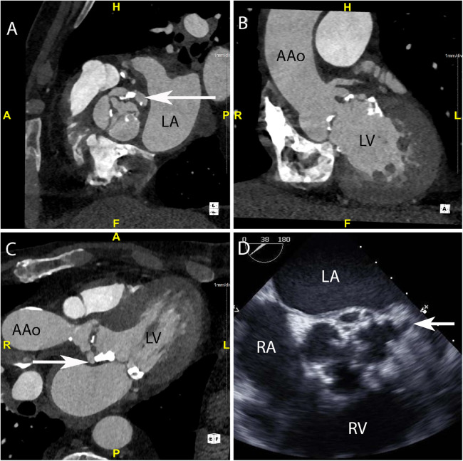
Infective Endocarditis (IE) remains a significant health challenge. Despite an increasing awareness, mortality is high and has remained largely unchanged over recent decades. Early diagnosis of IE is imperative and to assist clinicians several diagnostic criteria have been proposed. The best known are the Duke criteria. Originally published in 1994, these criteria have undergone significant modifications. This manuscript provides a timeline of the successive changes that have been made over the last 30 years. Changes which to a large degree have reflected both the evolving epidemiology of IE and the proliferation and increasing availability of advanced multi-modality imaging. Importantly, many of these changes now form part of societal guidelines for the diagnosis of IE. To provide validation for the incorporation of cardiac computed tomography (CT) in current guidelines, the manuscript demonstrates a spectrum of pictorial case studies that re-enforce the utility and growing importance of early cardiac CT in the diagnosis and treatment of suspected IE.

Keywords: Duke criteria; cardiac computed tomography (CT) imaging; infective endocarditis; pseudoaneursym; vegetations.


Conflict of interest statement

The authors declare that the research was conducted in the absence of any commercial or financial relationships that could be construed as a potential conflict of interest.

Figures

Figure 1
Figure 1
CASE 1 - A 75-year-old man presented feeling generally unwell with fever. Staphylococcus aureus was isolated from blood cultures (see text). LA, left atrium; LV, left ventricle; RA, right atrium; RV, right ventricle; AAo, ascending aorta.
Figure 2
Figure 2
CASE 2 - A 77-year-old man with a history of transcatheter aortic valve implantation (TAVI) presented with a sore throat, diarrhoea, and a temperature of 40°C. Staphylococcus aureus was isolated from blood cultures (see text). RA, right atrium; RVOT, right ventricular outflow tract; RV, right ventricle; LA, left atrium; LV, left ventricle; AAo, ascending aorta.
Figure 3
Figure 3
CASE 3 - A 79-year-old man presented with shortness of breath and fever. Blood cultures grew Staphylococcus aureus (see text). RA, right atrium; RV, right ventricle; LA, left atrium; LV, left ventricle; AAo, ascending aorta.
Figure 4
Figure 4
CASE 4 - A 79-year-old man with metallic mitral and aortic valve prostheses undergoing treatment for IE remained febrile and unwell despite appropriate guideline recommended antibiotic therapy (see text). RA, right atrium; RV, right ventricle; LA, left atrium; LV, left ventricle; AAo, ascending aorta.
Figure 5
Figure 5
CASE 5 - A 53-year-old man presented with fever, dyspnoea and confusion on a backgound history of intravenous drug use. Blood cultures grew Staphylococcus aureus (see text). RA, right atrium; RV, right ventricle; MPA, mean pulmonary artery; LA, left atrium; LV, left ventricle.
Figure 6
Figure 6
CASE 6 - A 71-year-old man presented with fever, confusion and fatigue. Enterococcus faecalis was isolated from blood cultures (see text). RA, right atrium; RVOT, right ventricular outflow tract; RV, right ventricle; MPA, mean pulmonary artery; LA, left atrium; LV, left ventricle; AAo, ascending aorta.
Figure 7
Figure 7
CASE 7 - A 69-year-old man presented with decompensated heart failure. Blood cultures were negative (see text). LAD, left anterior descending artery; LCX, left circumflex artery; RCA, right coronary artery; LA, left atrium; RA, right atrium; RVOT, right ventricular outflow tract.

References

    1. Mostaghim AS, Lo HYA, Khardori N. A retrospective epidemiologic study to define risk factors, microbiology, and clinical outcomes of infective endocarditis in a large tertiary-care teaching hospital. SAGE Open Med. (2017) 5:2050312117741772. 10.1177/2050312117741772 - DOI - PMC - PubMed
    1. Durack DT, Lukes AS, Bright DK. New criteria for diagnosis of infective endocarditis: utilization of specific echocardiographic findings. Duke endocarditis service. Am J Med. (1994) 96(3):200–9. 10.1016/0002-9343(94)90143-0 - DOI - PubMed
    1. Li JS, Sexton DJ, Mick N, Nettles R, Fowler VG, Jr., Ryan T, et al. Proposed modifications to the Duke criteria for the diagnosis of infective endocarditis. Clin Infect Dis. (2000) 30(4):633–8. 10.1086/313753 - DOI - PubMed
    1. Habib G, Lancellotti P, Antunes MJ, Bongiorni MG, Casalta JP, Del Zotti F, et al. 2015 ESC guidelines for the management of infective endocarditis: the task force for the management of infective endocarditis of the European Society of Cardiology (ESC). Endorsed by: European association for cardio-thoracic surgery (EACTS), the European association of nuclear medicine (EANM). Eur Heart J. (2015) 36(44):3075–128. 10.1093/eurheartj/ehv319 - DOI - PubMed
    1. Fowler VG, Durack DT, Selton-Suty C, Athan E, Bayer AS, Chamis AL, et al. The 2023 Duke-international society for cardiovascular infectious diseases criteria for infective endocarditis: updating the modified Duke criteria. Clin Infect Dis. (2023) 77(4):518–26. 10.1093/cid/ciad271 - DOI - PMC - PubMed

LinkOut - more resources