

Detection properties of indium-111 and IRDye800CW for intraoperative molecular imaging use across tissue phantom models
- PMID: 39310036
- PMCID: PMC11413652
- DOI: 10.1117/1.JBO.30.S1.S13705
Detection properties of indium-111 and IRDye800CW for intraoperative molecular imaging use across tissue phantom models
Abstract
Significance: Intraoperative molecular imaging (IMI) enables the detection and visualization of cancer tissue using targeted radioactive or fluorescent tracers. While IMI research has rapidly expanded, including the recent Food and Drug Administration approval of a targeted fluorophore, the limits of detection have not been well-defined.
Aim: The ability of widely available handheld intraoperative tools (Neoprobe and SPY-PHI) to measure gamma decay and fluorescence intensity from IMI tracers was assessed while varying characteristics of both the signal source and the intervening tissue or gelatin phantoms.
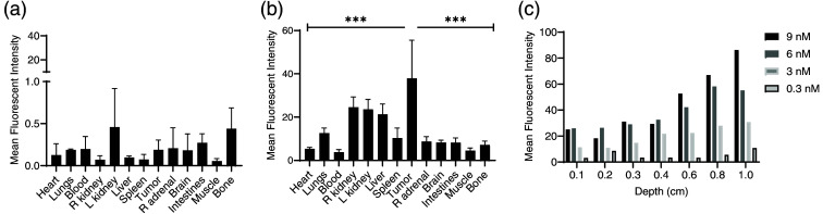
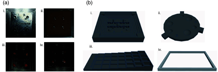
Approach: Gamma decay signal and fluorescence from tracer-bearing tumors (TBTs) and modifiable tumor-like inclusions (TLIs) were measured through increasing thicknesses of porcine tissue and gelatin in custom 3D-printed molds. TBTs buried beneath porcine tissue were used to simulate IMI-guided tumor resection.
Results: Gamma decay from TBTs and TLIs was detected through significantly thicker tissue and gelatin than fluorescence, with at least 5% of the maximum signal observed through up to 5 and 0.5 cm, respectively, depending on the overlying tissue type or gelatin.
Conclusions: We developed novel systems that can be fine-tuned to simulate variable tumor characteristics and tissue environments. These were used to evaluate the detection of fluorescent and gamma signals from IMI tracers and simulate IMI surgery.
Keywords: fluorescence; intraoperative molecular imaging; radioactivity; three-dimensional printing; tissue phantom; tumor detection.
© 2024 The Authors.
Figures


Similar articles
-
Prescription of Controlled Substances: Benefits and Risks.2025 Jul 6. In: StatPearls [Internet]. Treasure Island (FL): StatPearls Publishing; 2025 Jan–. 2025 Jul 6. In: StatPearls [Internet]. Treasure Island (FL): StatPearls Publishing; 2025 Jan–. PMID: 30726003 Free Books & Documents.
-
Systematic comparison of fluorescence imaging in the near-infrared and shortwave-infrared spectral range using clinical tumor samples containing cetuximab-IRDye800CW.J Biomed Opt. 2025 Jan;30(Suppl 1):S13708. doi: 10.1117/1.JBO.30.S1.S13708. Epub 2024 Nov 15. J Biomed Opt. 2025. PMID: 39553388 Free PMC article.
-
A Phase 2 Multicenter Clinical Trial of Intraoperative Molecular Imaging of Lung Cancer with a pH-Activatable Nanoprobe.Mol Imaging Biol. 2024 Aug;26(4):585-592. doi: 10.1007/s11307-024-01933-x. Epub 2024 Jul 11. Mol Imaging Biol. 2024. PMID: 38992245 Clinical Trial.
-
The Black Book of Psychotropic Dosing and Monitoring.Psychopharmacol Bull. 2024 Jul 8;54(3):8-59. Psychopharmacol Bull. 2024. PMID: 38993656 Free PMC article. Review.
-
Intraoperative imaging technology to maximise extent of resection for glioma: a network meta-analysis.Cochrane Database Syst Rev. 2021 Jan 4;1(1):CD013630. doi: 10.1002/14651858.CD013630.pub2. Cochrane Database Syst Rev. 2021. PMID: 33428222 Free PMC article.
Cited by
-
Introduction to the Special Issue on Molecular Guided Surgery.J Biomed Opt. 2025 Jan;30(Suppl 1):S13701. doi: 10.1117/1.JBO.30.S1.S13701. Epub 2025 May 7. J Biomed Opt. 2025. PMID: 40343094 Free PMC article.
References
-
- Sparber-Sauer M., et al. , “The significance of margins in pediatric non-rhabdomyosarcoma soft tissue sarcomas: consensus on surgical margin definition harmonization from the INternational Soft Tissue SaRcoma ConsorTium (INSTRuCT),” Cancer Med. 12(10), 11719–11730 (2023).10.1002/cam4.5671 - DOI - PMC - PubMed
Publication types
MeSH terms
Substances
Grants and funding
LinkOut - more resources
Full Text Sources
