

An Evasive Liver Mass in a Human Immunodeficiency Virus (HIV)-Positive Patient
- PMID: 39310052
- PMCID: PMC11415121
- DOI: 10.14309/crj.0000000000001481
An Evasive Liver Mass in a Human Immunodeficiency Virus (HIV)-Positive Patient
Abstract
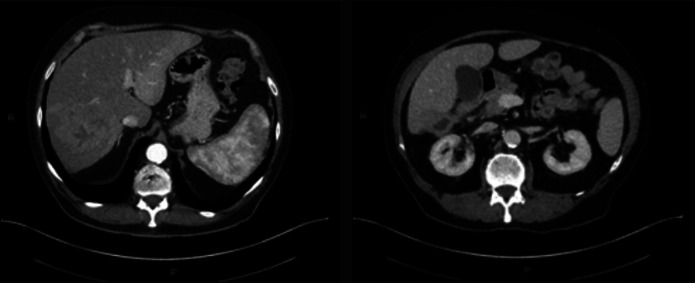
IgG4-related disease (IgG4-RD) is an autoimmune syndrome that is characterized by elevated levels of serum IgG4 and infiltration of various tissue types by IgG4 immunoreactive plasma cells. The IgG4-RD can result in systemic disease and the formation of inflammatory mass lesions, frequently addressed as pseudotumors. While IgG4-RD can manifest in various organs, liver involvement is rare, and because it is an immune-mediated inflammatory process, it is uncommon in patients who are immunocompromised. Furthermore, despite IgG4-RD responding well to immunosuppressive treatment, cases of spontaneous remission are exceedingly rare in the literature. In this report, we present the unique case of a self-resolving IgG4-RD lesion of the liver in a HIV positive patient.
Keywords: IgG4-related disease; immunosuppression; pseudotumor.
© 2024 The Author(s). Published by Wolters Kluwer Health, Inc. on behalf of The American College of Gastroenterology.
Figures


Similar articles
-
Immunoglobulin G4-related hepatobiliary disease.Semin Diagn Pathol. 2019 Nov;36(6):423-433. doi: 10.1053/j.semdp.2019.07.007. Epub 2019 Jul 24. Semin Diagn Pathol. 2019. PMID: 31358425 Review.
-
A Rare Case of Immunoglobulin G4-Related Disease Presenting With Coronary Artery Pseudotumor And Aneurysm.Eur J Case Rep Intern Med. 2023 Dec 6;11(1):004215. doi: 10.12890/2023_004215. eCollection 2024. Eur J Case Rep Intern Med. 2023. PMID: 38223278 Free PMC article.
-
Multivisceral IgG4-related disease presenting as recurrent massive gastrointestinal bleeding: a case report and literature review.BMC Gastroenterol. 2018 Sep 4;18(1):136. doi: 10.1186/s12876-018-0867-y. BMC Gastroenterol. 2018. PMID: 30180812 Free PMC article. Review.
-
Histopathology of IgG4-Related Autoimmune Hepatitis and IgG4-Related Hepatopathy in IgG4-Related Disease.Semin Liver Dis. 2016 Aug;36(3):229-41. doi: 10.1055/s-0036-1584320. Epub 2016 Jul 28. Semin Liver Dis. 2016. PMID: 27466793
-
Tumour of the orbit and pterygopalatine fossa: delayed recognition of possible IgG4-related disease.Contemp Oncol (Pozn). 2020;24(2):136-139. doi: 10.5114/wo.2020.97638. Epub 2020 Jul 3. Contemp Oncol (Pozn). 2020. PMID: 32774140 Free PMC article.
References
-
- Perugino CA, Stone JH. IgG4-related disease: An update on pathophysiology and implications for clinical care. Nat Rev Rheumatol. 2020;16(12):702–14. - PubMed
-
- Umehara H, Okazaki K, Kawa S, et al. . The 2020 revised comprehensive diagnostic (RCD) criteria for IgG4-RD. Mod Rheumatol. 2021;31(3):529–33. - PubMed
Publication types
LinkOut - more resources
Full Text Sources
Miscellaneous
