

Idiopathic Gingival Fibromatosis: Report of a Rare Case
- PMID: 39310417
- PMCID: PMC11415612
- DOI: 10.7759/cureus.67448
Idiopathic Gingival Fibromatosis: Report of a Rare Case
Abstract
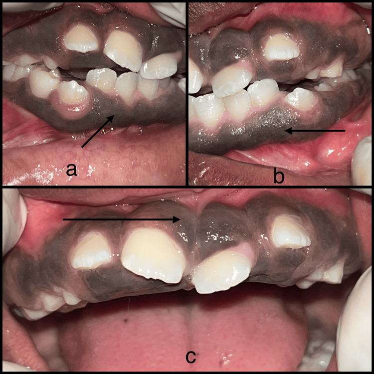
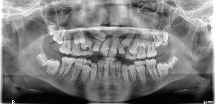
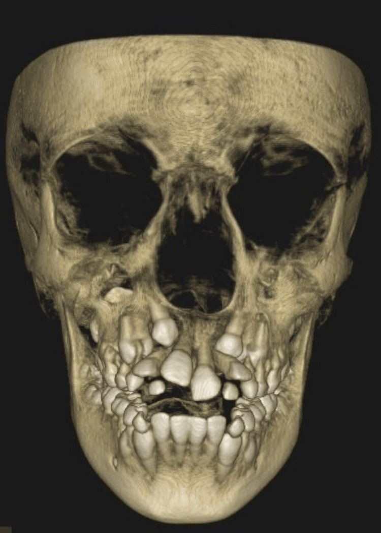
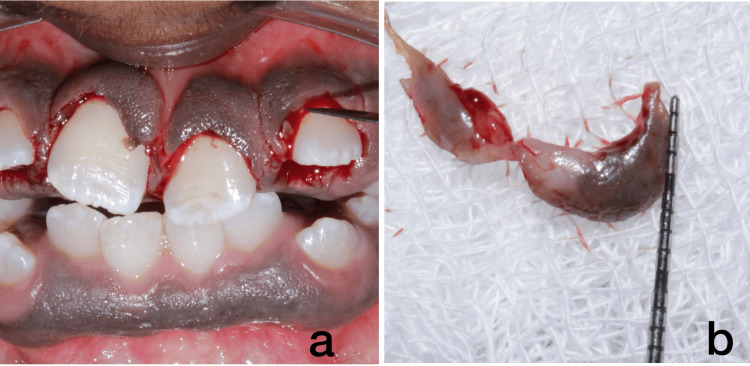
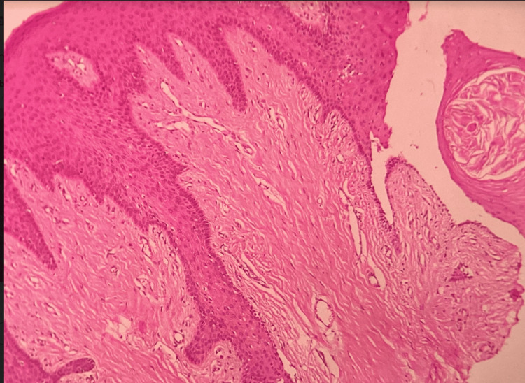
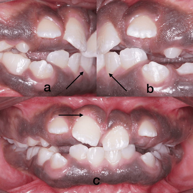
The progressive overgrowth of the gingiva is the hallmark of idiopathic gingival fibromatosis (IGF). Excess gingival tissue can obscure the crown of a tooth, resulting in spaces between teeth, displacement, retention of primary or permanent teeth, and difficulties with feeding, speaking, and appearance. The diagnosis and management of inherited gingival fibromatosis are the focus of this case report. A 12-year-old girl was referred from the Department of Orthodontics to Oral Medicine as a result of progressive gingival enlargement, which impeded orthodontic treatment for misaligned lower front teeth. The patient underwent a conservative periodontal treatment regimen that encompassed gingivectomy and debridement. The excised gingival tissues were submitted for histopathological examination. Tissue sections stained with hematoxylin and eosin showed connective tissue with dense bundles of collagen fibers and little inflammation. The patient was reviewed after three months, and advised of orthodontic management for further aesthetic correction. The findings indicated that the oral symptoms of gingival fibromatosis are influenced by the severity of the condition and the age at which it begins. Early intervention helps mitigate potential difficulties for younger individuals.
Keywords: gingival enlargement; gingival fibromatosis; gingival hyperplasia; gingival overgrowth; gingivectomy; idiopathic gingival fibromatosis.
Copyright © 2024, Shree Abiraami et al.
Conflict of interest statement
Human subjects: Consent was obtained or waived by all participants in this study. Conflicts of interest: In compliance with the ICMJE uniform disclosure form, all authors declare the following: Payment/services info: All authors have declared that no financial support was received from any organization for the submitted work. Financial relationships: All authors have declared that they have no financial relationships at present or within the previous three years with any organizations that might have an interest in the submitted work. Other relationships: All authors have declared that there are no other relationships or activities that could appear to have influenced the submitted work.
Figures
Similar articles
-
Hereditary gingival fibromatosis associated with generalized aggressive periodontitis: a case report.J Periodontol. 2004 May;75(5):770-8. doi: 10.1902/jop.2004.75.5.770. J Periodontol. 2004. PMID: 15212361
-
A Rare Case of non Syndromic Congenital Idiopathic Gingival Fibromatosis: Electrosurgical Management.J Clin Pediatr Dent. 2020 Sep 1;44(5):352-355. doi: 10.17796/1053-4625-44.5.10. J Clin Pediatr Dent. 2020. PMID: 33181851
-
Treatment of gingival fibromatosis associated with Zimmermann-Laband syndrome.J Periodontol. 2005 Sep;76(9):1559-62. doi: 10.1902/jop.2005.76.9.1559. J Periodontol. 2005. PMID: 16171447
-
Management of idiopathic gingival fibromatosis: report of a case and literature review.Pediatr Dent. 2011 Sep-Oct;33(5):431-6. Pediatr Dent. 2011. PMID: 22104713 Review.
-
Hereditary gingival fibromatosis in children: a systematic review of the literature.Clin Oral Investig. 2021 Jun;25(6):3599-3607. doi: 10.1007/s00784-020-03682-x. Epub 2020 Nov 13. Clin Oral Investig. 2021. PMID: 33188467
References
-
- Evidence of genetic heterogeneity for hereditary gingival fibromatosis. Hart TC, Pallos D, Bozzo L, Almeida OP, Marazita ML, O'Connell JR, Cortelli JR. J Dent Res. 2000;79:1758–1764. - PubMed
Publication types
LinkOut - more resources
Full Text Sources