

Correlation Between Radiological Changes of the Temporomandibular Joint and Upper Cervical Vertebrae in Degenerative Joint Disease: A Cone-Beam Computed Tomography-Based Analytical Study
- PMID: 39310450
- PMCID: PMC11416149
- DOI: 10.7759/cureus.67518
Correlation Between Radiological Changes of the Temporomandibular Joint and Upper Cervical Vertebrae in Degenerative Joint Disease: A Cone-Beam Computed Tomography-Based Analytical Study
Abstract
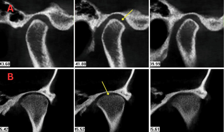
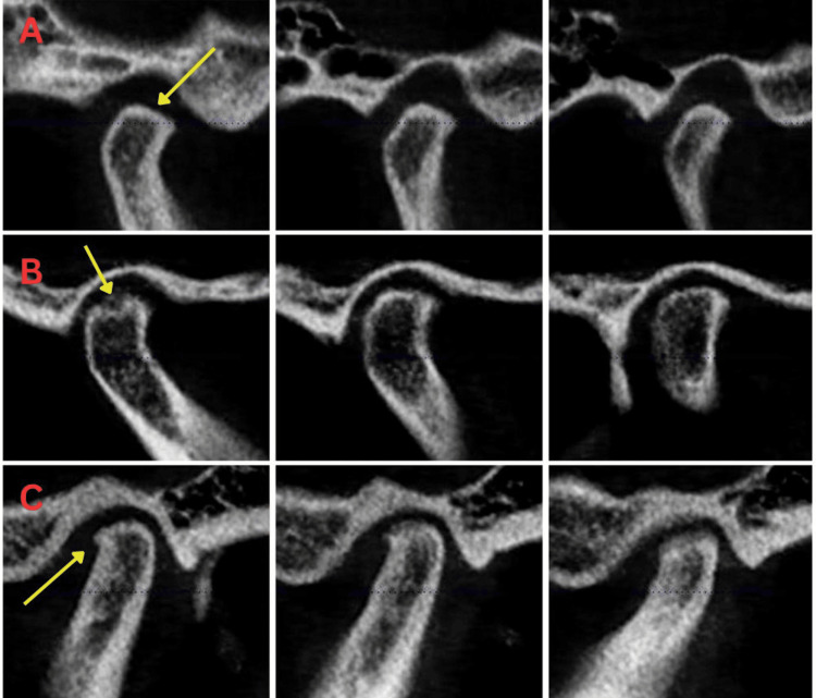
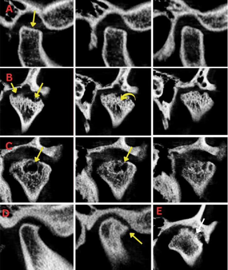
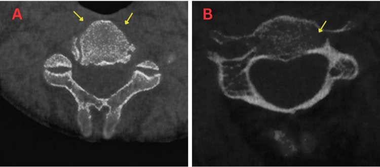
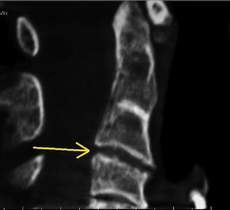
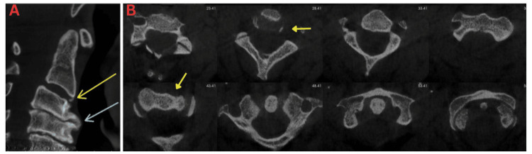
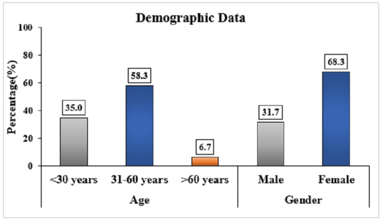
Objectives This study was conducted to assess the radiological changes of the temporomandibular joint (TMJ) and cervical vertebrae individually and their correlation in degenerative joint disease (DJD) using a cone-beam computed tomography (CBCT)-based approach. Methodology The study employed a cross-sectional, analytical retrospective design, analyzing one-year data. CBCT scans of 60 patients (120 TMJs) were assessed for degenerative changes using standardized imaging parameters. Eligibility criteria included full field-of-view CBCT scans, excluding those with craniofacial anomalies or prior orthodontic treatment. Radiological assessments of TMJs and cervical vertebrae were conducted by experienced radiologists using the Anjos Pontual method and novel grading system (TMJ Spine Degenerative Severity Index). Results The study included 60 CBCT scans (120 joints), with 31.7% males and 68.3% females. Participants were predominantly aged 31-60 years (58.3%). DJD findings for the right TMJ showed grade 1 changes in 55.0% and grade 2 in 31.7%, while the left TMJ had 46.7% grade 1 and 35.0% grade 2 changes. A strong positive correlation (0.704) was found between bilateral TMJ and cervical vertebrae changes. Age correlated significantly with TMJ alterations but not with cervical vertebrae changes. Conclusion This study demonstrated that there exists a positive association between the radiological changes of TMJ and cervical vertebrae in DJD with age, which can be detected in mild stage of severity on CBCT and can be of use in clinical correlation and application of optimal interventions ensuring better prognosis.
Keywords: cone-beam computed tomography (cbct); degenerative joint disease; diagnostic ct imaging; severity of disease; temporomandibular joint (tmj); upper cervical spine.
Copyright © 2024, Nayak et al.
Conflict of interest statement
Human subjects: Consent was obtained or waived by all participants in this study. Institutional Ethics Committee, Terna Dental College issued approval TDC/EC/22/2023. Animal subjects: All authors have confirmed that this study did not involve animal subjects or tissue. Conflicts of interest: In compliance with the ICMJE uniform disclosure form, all authors declare the following: Payment/services info: All authors have declared that no financial support was received from any organization for the submitted work. Financial relationships: All authors have declared that they have no financial relationships at present or within the previous three years with any organizations that might have an interest in the submitted work. Intellectual property info: The novel classification system, "CBCT-Based Grading System for Severity in Degenerative Joint Disease of TMJ and Upper Cervical Spine" is granted approval and registered with Ref. No. L-152146/2024 under the Copyright Office, Intellectual Property Rights, Government of India. Other relationships: All authors have declared that there are no other relationships or activities that could appear to have influenced the submitted work.
Figures
Similar articles
-
Limited implication of initial bone scintigraphy on long-term condylar bone change in temporomandibular disorders-Comparison with cone beam computed tomography at 1 year.J Oral Rehabil. 2021 Aug;48(8):880-890. doi: 10.1111/joor.13209. Epub 2021 Jun 8. J Oral Rehabil. 2021. PMID: 34032306
-
Degenerative Joint Disease of the Upper Cervical Spines: A Cone Beam Computed Tomography Study.Niger J Clin Pract. 2020 Dec;23(12):1667-1672. doi: 10.4103/njcp.njcp_628_19. Niger J Clin Pract. 2020. PMID: 33355819
-
Association of radiographic and clinical findings in patients with temporomandibular joints osseous alteration.Clin Oral Investig. 2020 Jan;24(1):221-227. doi: 10.1007/s00784-019-02945-6. Epub 2019 May 11. Clin Oral Investig. 2020. PMID: 31079244
-
A Systematic Review on the Association Between Clinical Symptoms and CBCT Findings in Symptomatic TMJ Degenerative Joint Disease.J Oral Facial Pain Headache. 2021 Nov-Dec;35(4):332-345. doi: 10.11607/ofph.2953. J Oral Facial Pain Headache. 2021. PMID: 34990502
-
What is the image appearance of juvenile idiopathic arthritis in MRI, CT, and CBCT of TMJ? A systematic review.Clin Oral Investig. 2023 May;27(5):2321-2333. doi: 10.1007/s00784-022-04828-9. Epub 2022 Dec 14. Clin Oral Investig. 2023. PMID: 36515761
Cited by
-
Assessment of degenerative changes in the atlanto-odontoid joint using cone-beam computed tomography (CBCT) imaging.PeerJ. 2025 Jun 10;13:e19569. doi: 10.7717/peerj.19569. eCollection 2025. PeerJ. 2025. PMID: 40520641 Free PMC article.
References
-
- Knee orthopedics as a template for the temporomandibular joint. Bielajew BJ, Donahue RP, Espinosa MG, et al. https://www.sciencedirect.com/science/article/pii/S2666379121000574. Cell Rep Med. 2021;2:100241. - PMC - PubMed
-
- Evaluation of the severity of temporomandibular joint osteoarthritic changes related to age using cone beam computed tomography. Alexiou K, Stamatakis H, Tsiklakis K. Dentomaxillofac Radiol. 2009;38:141–147. - PubMed
-
- Degenerative joint disease of the upper cervical spines: a cone beam computed tomography study. Khalifa HM, Alamoudi AA, Jan AM, Jadu FM. Niger J Clin Pract. 2020;23:1667–1672. - PubMed
LinkOut - more resources
Full Text Sources