Insight Into Uncommon Territory: Exploring Internuclear Ophthalmoplegia in Artery of Percheron Infarct
- PMID: 39310574
- PMCID: PMC11416046
- DOI: 10.7759/cureus.67485
Insight Into Uncommon Territory: Exploring Internuclear Ophthalmoplegia in Artery of Percheron Infarct
Abstract
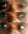
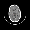
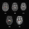
Internuclear ophthalmoplegia (INO), a neurological disorder is characterized by horizontal gaze palsy because of a lesion in the medial longitudinal fasciculus, a neural pathway that is mainly responsible for coordinating the movements of the eye. INO presents with diplopia and impaired adduction of the affected eye, accompanied by abducting eye nystagmus. The condition also arises from different etiologies which include multiple sclerosis, encephalitis, Lyme disease, HIV, and herpes zoster. Artery of Percheron (AOP) infarction is a subtype of bilateral thalamic infarction that poses a unique form of diagnostic perplexity due to its varied and often non-specific clinical manifestations such as altered responsiveness, memory disturbances, and oculomotor deficits. Here we discuss a 53-year-old female who presented with INO in the context of an AOP infarct. Under this context, the clinical finding includes some paradigms like nystagmus, anisocoria, and bilateral ptosis. Magnetic resonance imaging confirmed an acute infarct in the AOP territory, which supplies the rostral midbrain and paramedian thalami. This case emphasizes the critical importance of meticulous clinical evaluation and the utilization of advanced imaging techniques in diagnosing rare stroke syndromes like AOP infarction. Management of the patient included dual antiplatelet therapy to prevent further thromboembolic events and supportive care to address the immediate neurological deficits. Early recognition and prompt treatment are crucial for better patient outcomes. Long-term management focuses on the secondary prevention of stroke through lifestyle modifications, medical therapy, and regular monitoring. Education on uncommon stroke syndromes and continued research are essential for enhancing the accuracy of diagnosis and efficacy of treatment which ultimately leads to better patient care and prognosis.
Keywords: artery of percheron; internuclear ophthalmoplegia; medial longitudinal fasciculus; stroke syndromes; thalamic infarction.
Copyright © 2024, Jayakumar et al.
Conflict of interest statement
Human subjects: Consent was obtained or waived by all participants in this study. Conflicts of interest: In compliance with the ICMJE uniform disclosure form, all authors declare the following: Payment/services info: All authors have declared that no financial support was received from any organization for the submitted work. Financial relationships: All authors have declared that they have no financial relationships at present or within the previous three years with any organizations that might have an interest in the submitted work. Other relationships: All authors have declared that there are no other relationships or activities that could appear to have influenced the submitted work.
Figures


References
-
- Clinical insights into internuclear ophthalmoplegia: a case report. Rahmadiansyah MF, Hidayat M. Bioscientia Medicina: Journal of Biomedicine and Translational Research. 2023;7:3789–3794.
-
- Unilateral internuclear ophthalmoplegia due to microvascular pontine infarction. Hossain S, Guda TL, Chowdhury F, Hossain MS. Anwer Khan Modern Medical College Journal. 2018;9:71–74.
-
- Artery of percheron infarct: a rare presentation of acute ischemic stroke in a high-risk antiphospholipid syndrome patient. Sreethish Sasi, Ashraf Ahmed, Wajiha Yousuf, et al. Case Reports in Acute Medicine. 2020;3:46–52.
-
- A clinical study of artery of Percheron infarction. Chiang YK, Ling YH, Chang FC, Fuh JL. J Chin Med Assoc. 2022;85:1098–1100. - PubMed
Publication types
LinkOut - more resources
Full Text Sources
