

BIOFILM AND HISTOPATHOLOGICAL GRADING OF MAXILLARY SINUS MUCOSA IN PATIENTS WITH ANTROCHOANAL POLYPS
- PMID: 39310685
- PMCID: PMC11414000
- DOI: 10.20471/acc.2023.62.03.2
BIOFILM AND HISTOPATHOLOGICAL GRADING OF MAXILLARY SINUS MUCOSA IN PATIENTS WITH ANTROCHOANAL POLYPS
Abstract
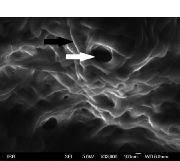
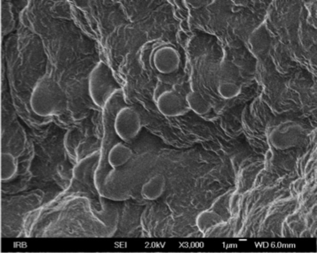
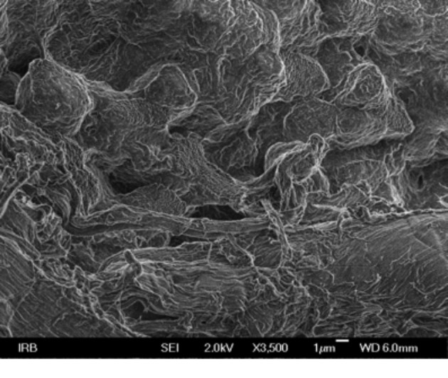
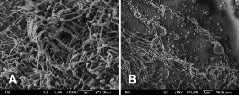
The aim of this cross-sectional study was to determine the signs of biofilm in the maxillary sinus of patients with antrochoanal polyps (ACP), and status of the mucosa on which the biofilm occurred. Mucosal samples from maxillary sinus in 40 ACP patients who underwent endoscopic sinus surgery were analyzed histopathologically and by scanning electron microscopy. Results were compared with maxillary mucosa samples of 40 patients without endoscopic and radiological signs of sinus disease. The existence of biofilm and its relation to the degree of histopathological changes according to Terrier classification of chronic mucosal inflammation of maxillary sinus were statistically analyzed. Biofilm was detected in 23 of 40 (57.5%) ACP patients; the incidence was significantly lower in the control group (2/40, 5%). Biofilm was not found in type 1 mucosa according to Terrier classification. In conclusion, biofilm showed a significant incidence in the maxillary sinus mucosa of ACP patients (57.5%). Occasionally, biofilm can be found in patients with no signs of sinus disease, but not on histologically normal mucosa. Results of this study support the theory that biofilm formation does not represent the initial stage of the inflammatory process.
Keywords: Antrochoanal polyp; Biofilm; Maxillary sinus.
Sestre Milosrdnice University Hospital.
Figures
Similar articles
-
Biofilm in nasal polyps.Rhinology. 2008 Dec;46(4):302-7. Rhinology. 2008. PMID: 19146001
-
The analysis of the maxillary sinus volumes and the nasal septal deviation in patients with antrochoanal polyps.Eur Arch Otorhinolaryngol. 2015 Nov;272(11):3347-52. doi: 10.1007/s00405-014-3460-1. Epub 2014 Dec 23. Eur Arch Otorhinolaryngol. 2015. PMID: 25534286
-
Endoscopic management of paediatric antrochoanal polyp: our experience.Acta Otorhinolaryngol Ital. 2013 Apr;33(2):107-11. Acta Otorhinolaryngol Ital. 2013. PMID: 23853401 Free PMC article.
-
Antrochoanal polyp arising from benign pseudocyst of maxillary antrum.J Indian Soc Pedod Prev Dent. 2017 Jul-Sep;35(3):275-278. doi: 10.4103/JISPPD.JISPPD_153_16. J Indian Soc Pedod Prev Dent. 2017. PMID: 28762356 Review.
-
The antrochoanal polyp.Rhinology. 2004 Dec;42(4):178-82. Rhinology. 2004. PMID: 15626248 Review.
References
MeSH terms
LinkOut - more resources
Full Text Sources