. 2024 Aug 30;27(9):110846.
doi: 10.1016/j.isci.2024.110846. eCollection 2024 Sep 20.

AXL and SHC1 confer crizotinib resistance in patient-derived xenograft model of ALK-driven lung cancer

Affiliations

AXL and SHC1 confer crizotinib resistance in patient-derived xenograft model of ALK-driven lung cancer

Yerong Hu et al. iScience. .

Abstract

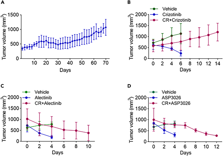
Anaplastic lymphoma kinase (ALK) inhibitor crizotinib has dramatic effect in non-small cell lung cancer patients with ALK rearrangement. However, most patients eventually develop resistance. To discover therapeutic targets to overcome crizotinib resistance (CR), we generated patient-derived xenograft CR mice and subjected them to phosphorylation profiling, together with CR mice treated with ASP3026 or alectinib. We identified 100 proteins with different phosphorylation status in CR mice. Among them, AXL phosphorylation was increased in CR mice, which could not be reversed by ASP3026 or alectinib. Importantly, the combined treatment of crizotinib and AXL inhibitor in CR mice significantly inhibited tumor growth, compared to crizotinib alone. We also found that SHC1 phosphorylation was increased in CR mice and SHC1 knockdown sensitized ALK-driven cells to crizotinib. Our study provides a new view of signaling pathways leading to CR, suggesting AXL and SHC1 as potential targets for combination therapy to overcome CR.

Keywords: Cancer; Drugs; Molecular biology.


Conflict of interest statement

The authors declare no competing interests.

Figures

None
Graphical abstract
Figure 1
Figure 1
ALK protein expression and the type of EML4-ALK variant in tumor tissues from NSCLC patients and PDX mice (A) Western blot analysis of protein lysates from tumor tissues of representative NSCLC patients using antibody against ALK and GAPDH was used as a loading control. Positions of EML4-ALK variants are indicated. (B) Sequencing analysis of EML4-ALK variants in tumor tissues from representative NSCLC patients. ALK fragments were amplified using primer pairs listed in Table S1. The sequence characteristics of different EML4-ALK variants are indicated by arrows, which mark the starting point of exon 20 of ALK (V1: EML4-ALK variant 1, V2: EML4-ALK variant 2, V3: EML4-ALK variant 3). (C) RT-PCR analysis of EML4-ALK (variant 3) expression in tumor tissues from the PDX mice (P1–P5) and donor patient (T5, P0). The PCR primers used to amplify the EML4 and ALK cDNA are listed in Table S1. Amplification of GAPDH was used as a qualitative control of the cDNA samples. (D) Immunohistochemical staining of tumor tissues from the patient (T5, P0) and PDX mice (P1–P5) with ALK antibody (ALK (D5F3) XP Rabbit mAb). The ALK protein was stained in red.
Figure 2
Figure 2
The effects of different ALK inhibitors on tumor growth in crizotinib-resistant PDX mice (A) The tumor growth curve of the last round of crizotinib treatment (100 mg/kg/day) in 14 crizotinib-resistant PDX mice. Tumor sizes were measured every other day. (B) The tumor growth curves of crizotinib-resistant PDX mice further treated with crizotinib (100 mg/kg/day) and crizotinib-sensitive PDX mice treated with crizotinib or vehicle (N = 4). Tumor volumes were measured every other day. (C) The tumor growth curves of crizotinib-resistant PDX mice when treated with alectinib (60 mg/kg/day) and crizotinib-sensitive PDX mice when treated with alectinib and vehicle (N = 4). (D) The tumor growth curves of crizotinib-resistant PDX mice when treated with ASP3026 (100 mg/kg/day) and crizotinib-sensitive PDX mice when treated with ASP3026 and vehicle (N = 4).
Figure 3
Figure 3
PhosphoScan profiling of tumor tissues from crizotinib-resistant PDX mouse model (A) Immunoblotting with P-Tyr-100 of crizotinib-resistant PDX tumor tissues treated with the indicated inhibitors and crizotinib-sensitive PDX tumor tissues treated with crizotinib or vehicle (#1 and #2 represent tumor tissues from two different mice in the same group; CS, crizotinib-sensitive; CR, crizotinib-resistant; Con, control; Criz, crizotinib; Ale, alectinib; ASP, ASP3026). (B) PhosphoScan analysis of the proteins whose phosphorylation levels were increased by more than 2.5-folds (Table S3) in crizotinib-resistant PDX tumors compared to crizotinib-sensitive PDX tumors by protein type (N = 2). (C) Confirmation of MS/MS results by immunoblotting with indicated antibodies. β-actin was used as the loading control (#1and #2 represent tumor tissues from two different mice in the same group; CS, crizotinib-sensitive; CR, crizotinib-resistant; Con, control; Criz, crizotinib; Ale, alectinib; ASP, ASP3026).
Figure 4
Figure 4
The cellular signaling networks involved in crizotinib resistance (N = 2) (A) Venn diagram of 100 proteins whose phosphorylation levels were increased by more than 2.5-folds in crizotinib-resistant PDX tumors compared to crizotinib-sensitive PDX tumors. The numbers of proteins whose phosphorylation were reversed by alectinib or ASP3026 are shown. (B) 20 proteins whose phosphorylation in crizotinib-resistant tumors were reversed by both ASP3026 and alectinib (CS, crizotinib-sensitive; CR, crizotinib-resistant; Ale, alectinib; ASP, ASP3026). (C) Top 30 proteins whose increased phosphorylation in crizotinib-resistant tumors were affected by ASP3026 or alectinib (CS, crizotinib-sensitive; CR, crizotinib-resistant; Ale, alectinib; ASP, ASP3026).
Figure 5
Figure 5
The effects of bemcentinib and siSHC1 on NCI-3122 cells and crizotinib-resistant PDX tumors (A) Cell viability assays of NCI-H3122 and NCI-H3122-CR23 cells treated with crizotinib (Criz) for 72 h. The data are presented as mean ± SD from three independent experiments. (B) Immunoblotting with P-Tyr-100 of NCI-H3122 and NCI-H3122-CR23 cells treated with 1 μmol/L crizotinib for 4 h. Tubulin was used as the loading control. (C) Sequencing analysis of EML4-ALK showing a kinase domain heterozygous mutation F1174C in NCI-H3122-CR23. TTC in exon 23 of the ALK kinase region is replaced by TGC. (D) Immunoblotting of AXL in NCI-H3122-CR23 cells treated with different concentrations of bemcentinib (Bemcen) and NCI-H3122 with Veh (vehicle) for 72 h. Cell lysates were probed with the indicated antibodies. Tubulin was used as the loading control. (E) The viability of NCI-H3122-CR23 measured by MTS assay after treatment of bemcentinib (Bemcen) in combination with crizotinib (Criz) for 72 h. Results are representative of three independent experiments. The data are presented as mean ± SD. ∗∗p ≤ 0.01. ∗∗∗p ≤ 0.001. (F) The inhibition effect by the combined treatment of crizotinib and bemcentinib in the crizotinib-resistant PDX mouse model. Tumor volume fold changes and TIR (tumor inhibition ratio) were indicated with drugs. TIR was calculated after the 25-day treatment. ∗p < 0.05. ∗∗p < 0.01. ∗∗∗p ≤ 0.001. (G) Immunoblotting of AXL and ALK in crizotinib-resistant PDX tumors treated with drugs indicated under the same conditions as in Figure 5F. Tissue lysates were probed with the indicated antibodies. Tubulin was used as the loading control. Veh, Vehicle; Criz, crizotinib; Criz and Bemcen, crizotinib and bemcentinib. (H) Immunoblotting of NCI-H3122 cells treated with 1 μmol/L crizotinib for 4 h. Cell lysates were probed with the indicated antibodies. Veh, vehicle; Criz, Crizotinib. (I) SHC1 mRNA levels in NCI-H3122 cells transfected with SHC1 siRNAs (siSHC1) for 72 h. Cells mRNA were amplificated with the primers presented in Table S1. Cont siRNA, negative control virus vector; #1 and #2 were two different interfering sequences of SHC1 mRNA. (J) Immunoblotting of NCI-H3122 cells transfected with SHC1 siRNAs (siSHC1) #2 for 72 h; cell lysates were probed with the indicated antibodies. (K) Cell viability assay of NCI-H3122 cells. NCI-H3122 cells were transfected with SHC1 siRNAs (siSHC1) #2 for 72 h and then treated with the indicated concentration of crizotinib for 72 h. Control cells were transfected with scrambled siRNA. Results are representative of three independent experiments. The data are presented as mean ± SD. Cont siRNA, negative control virus vector; Criz, crizotinib. ∗∗p < 0.01. ∗∗∗∗p = 0.000.

References

    1. Soda M., Choi Y.L., Enomoto M., Takada S., Yamashita Y., Ishikawa S., Fujiwara S.i., Watanabe H., Kurashina K., Hatanaka H., et al. Identification of the transforming EML4-ALK fusion gene in non-small-cell lung cancer. Nature. 2007;448:561–566. doi: 10.1038/nature05945. - DOI - PubMed
    1. Shaw A.T., Kim D.W., Nakagawa K., Seto T., Crinó L., Ahn M.J., De Pas T., Besse B., Solomon B.J., Blackhall F., et al. Crizotinib versus chemotherapy in advanced ALK-positive lung cancer. N. Engl. J. Med. 2013;368:2385–2394. doi: 10.1056/NEJMoa1214886. - DOI - PubMed
    1. Solomon B.J., Mok T., Kim D.W., Wu Y.L., Nakagawa K., Mekhail T., Felip E., Cappuzzo F., Paolini J., Usari T., et al. First-line crizotinib versus chemotherapy in ALK-positive lung cancer. N. Engl. J. Med. 2014;371:2167–2177. doi: 10.1056/NEJMoa1408440. - DOI - PubMed
    1. Shaw A.T., Engelman J.A. ALK in lung cancer: past, present, and future. J. Clin. Oncol. 2013;31:1105–1111. doi: 10.1200/JCO.2012.44.5353. - DOI - PMC - PubMed
    1. Choi Y.L., Soda M., Yamashita Y., Ueno T., Takashima J., Nakajima T., Yatabe Y., Takeuchi K., Hamada T., Haruta H., et al. EML4-ALK mutations in lung cancer that confer resistance to ALK inhibitors. N. Engl. J. Med. 2010;363:1734–1739. doi: 10.1056/NEJMoa1007478. - DOI - PubMed

LinkOut - more resources