Case Reports
. 2023 Aug 3;32(3):266-270.
doi: 10.4103/jmu.jmu_103_22. eCollection 2024 Jul-Sep.

Uterine Arteriovenous Malformations

Affiliations
Case Reports

Uterine Arteriovenous Malformations

Martina Bartolone et al. J Med Ultrasound. .

Abstract

Uterine arteriovenous malformations (AVMs) are an abnormal presence of shunts between myometrial arteries and veins within the myometrium that usually occurs after a traumatic event on the uterus and it is often diagnosed after a miscarriage. In this case report, we propone the case of a woman, gravida 3 para 2, admitted at the emergency department presenting deep vaginal bleeding and suspicion of incomplete miscarriage at 11 weeks of pregnancy. The suspect of AVM was made with noninvasive procedure; transvaginal ultrasound examination with the advantage of color Doppler showed a myometrial hypervascular lesion of the posterior wall. Pulsed Doppler permitted the waveform analysis of uterine arteries and three-dimensional sonography with color Doppler and reconstructions clearly showed dilated ad tortuous blood vessels within the contest of the myometrium. Magnetic resonance angiography showed multiple tubular structures with tortuous appearance that confirmed the suspicion of AVM. Uterine artery embolization was performed of the right uterine artery. One month after uterine embolization, the ultrasound control confirmed the complete resolution of the AVM.

Keywords: Embolization; magnetic resonance; ultrasound; uterine arteriovenous malformation; vaginal bleeding.


Conflict of interest statement

There are no conflicts of interest.

Figures

Figure 1
Figure 1
Grayscale bidimensional ultrasound image showing longitudinal view of uterine body with anechoic area suspect for incomplete miscarriage
Figure 2
Figure 2
Grayscale bidimensional sonogram showing longitudinal view of uterine body with an inhomogeneous endometrium with multiple anechoic spaces. A free fluid of the cul the sac of Douglas is also visualized
Figure 3
Figure 3
Color Doppler ultrasound sagittal view of the uterus with turbulent flow and aliasing creating a mosaic pattern
Figure 4
Figure 4
Color mosaic pattern of the myometrium analyzed with High Definition Power Doppler
Figure 5
Figure 5
Spectral analysis revealing low-resistance index suggesting arteriovenous malformation
Figure 6
Figure 6
(a and b) Three-dimensional power Doppler ultrasound reconstruction of the uterus using a glass body technique to differentiate tissue and vessels. This method reveals a hypervascular lesion in the myometrium
Figure 7
Figure 7
(a and b) Surface-rendered reconstruction showing irregular vessels of arteriovenous malformation in bicolor and monochrome Three-dimensional high-definition flow
Figure 8
Figure 8
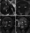
Sagittal and axial T2-weighted sequences (a and b) showing an enlarged uterus with hypointense lesion at the right lateral wall extending into the uterine cavity. After administration of intravenous contrast, marked enhancement is present in the early arterial phase (c and d). Arrows indicate myometrial and endometrial vessels
Figure 9
Figure 9
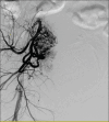
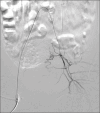
Images of the contrast material injected into the right uterine artery that revealed the enhanced myometrial vascularity/ arteriovenous malformation before (image A and B) and after the uterine artery embolization (image C)
Figure 10
Figure 10
Images of the contrast material injected into the right uterine artery that revealed the enhanced myometrial vascularity/arteriovenous malformation before artery embolization
Figure 11
Figure 11
Images of the contrast material injected into the right uterine artery that revealed the enhanced myometrial vascularity/arteriovenous malformation after the uterine artery embolization
Figure 12
Figure 12
Transvaginal ultrasound image 1 month after embolization
Figure 13
Figure 13
Magnetic resonance performed 1 month after treatment. Sagittal plane on T2 (a) and on T1 (b) after contrast enhancement. Arrows indicate endometrium

References

    1. Diwan RV, Brennan JN, Selim MA, McGrew TL, Rashad FA, Rustia MU, et al. Sonographic diagnosis of arteriovenous malformation of the uterus and pelvis. J Clin Ultrasound. 1983;11:295–8. - PubMed
    1. Vogelzang RL, Nemcek AA, Jr, Skrtic Z, Gorrell J, Lurain JR. Uterine arteriovenous malformations: Primary treatment with therapeutic embolization. J Vasc Interv Radiol. 1991;2:517–22. - PubMed
    1. Timor-Tritsch IE, Haynes MC, Monteagudo A, Khatib N, Kovács S. Ultrasound diagnosis and management of acquired uterine enhanced myometrial vascularity/arteriovenous malformations. Am J Obstet Gynecol. 2016;214:731.e1–731.e10. - PubMed
    1. Polat P, Suma S, Kantarcý M, Alper F, Levent A. Color Doppler US in the evaluation of uterine vascular abnormalities. Radiographics. 2002;22:47–53. - PubMed
    1. Yoon DJ, Jones M, Taani JA, Buhimschi C, Dowell JD. A systematic review of acquired uterine arteriovenous malformations: Pathophysiology, diagnosis, and transcatheter treatment. AJP Rep. 2016;6:e6–14. - PMC - PubMed

Publication types

LinkOut - more resources