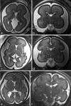
Prenatal Ultrasound and Magnetic Resonance Findings of Glutaric Acidemia Type 1 and Its Challenges in Prenatal Diagnosis
- PMID: 39310876
- PMCID: PMC11414954
- DOI: 10.4103/jmu.jmu_63_24
Prenatal Ultrasound and Magnetic Resonance Findings of Glutaric Acidemia Type 1 and Its Challenges in Prenatal Diagnosis
Abstract
Glutaric acidemia type 1 (GA1) presents unique challenges in prenatal diagnosis, especially in cases with no family history. This review article aims to review and present the prenatal ultrasound and magnetic resonance findings of GA1 and consolidate key insights into the difficulties associated with GA1 prenatal diagnosis and the neuroimaging features that require careful differentiation during the diagnostic process.
Keywords: Fetal magnetic resonance imaging; Sylvian fissure; glutaric acidemia type 1; prenatal ultrasound.
Copyright: © 2024 Journal of Medical Ultrasound.
Conflict of interest statement
Dr. Tung-Yao Chang, an editorial board member at Journal of Medical Ultrasound, had no role in the peer review process of or decision to publish this article. The other authors declared no conflicts of interest in writing this paper.
Figures


Similar articles
-
Prenatal diagnosis of glutaric acidemia type 2 with the use of exome sequencing - an up-to-date review and new case report.Ginekol Pol. 2021;92(1):51-56. doi: 10.5603/GP.a2020.0190. Epub 2021 Jan 15. Ginekol Pol. 2021. PMID: 33448012
-
Occurrence of subdural hematomas in Dutch glutaric aciduria type 1 patients.Eur J Pediatr. 2016 Jul;175(7):1001-6. doi: 10.1007/s00431-016-2734-6. Epub 2016 May 31. Eur J Pediatr. 2016. PMID: 27246831 Free PMC article.
-
Biochemical and molecular features of chinese patients with glutaric acidemia type 1 from Fujian Province, southeastern China.Orphanet J Rare Dis. 2023 Jul 26;18(1):215. doi: 10.1186/s13023-023-02833-z. Orphanet J Rare Dis. 2023. PMID: 37496092 Free PMC article.
-
[Mass spectrometry combined with gene analysis for prenatal diagnosis of glutaric acidemia type Ⅰ].Zhonghua Er Ke Za Zhi. 2017 Jul 2;55(7):539-543. doi: 10.3760/cma.j.issn.0578-1310.2017.07.014. Zhonghua Er Ke Za Zhi. 2017. PMID: 28728265 Chinese.
-
Glutaric Acidemia, Pathogenesis and Nutritional Therapy.Front Nutr. 2021 Dec 15;8:704984. doi: 10.3389/fnut.2021.704984. eCollection 2021. Front Nutr. 2021. PMID: 34977106 Free PMC article. Review.
References
-
- Boy N, Mengler K, Thimm E, Schiergens KA, Marquardt T, Weinhold N, et al. Newborn screening: A disease-changing intervention for Glutaric aciduria type 1. Ann Neurol. 2018;83:970–9. - PubMed
-
- Tsai FC, Lee HJ, Wang AG, Hsieh SC, Lu YH, Lee MC, et al. Experiences during newborn screening for Glutaric aciduria type 1: Diagnosis, treatment, genotype, phenotype, and outcomes. J Chin Med Assoc. 2017;80:253–61. - PubMed
-
- Boy N, Mühlhausen C, Maier EM, Ballhausen D, Baumgartner MR, Beblo S, et al. Recommendations for diagnosing and managing individuals with Glutaric aciduria type 1: Third revision. J Inherit Metab Dis. 2023;46:482–519. - PubMed
-
- Schuurmans IM, Dimitrov B, Schröter J, Ribes A, de la Fuente RP, Zamora B, et al. Exploring genotype-phenotype correlations in Glutaric aciduria type 1. J Inherit Metab Dis. 2023;46:371–90. - PubMed
-
- Wajner M, Amaral AU, Leipnitz G, Seminotti B. Pathogenesis of brain damage in Glutaric acidemia type I: Lessons from the genetic mice model. Int J Dev Neurosci. 2019;78:215–21. - PubMed
Publication types
LinkOut - more resources
Full Text Sources
