. 2024 Aug 30;13(3):71.
doi: 10.3390/antib13030071.

Functional Activity of Cytokine-Induced Killer Cells Enhanced by CAR-CD19 Modification or by Soluble Bispecific Antibody Blinatumomab

Affiliations

Functional Activity of Cytokine-Induced Killer Cells Enhanced by CAR-CD19 Modification or by Soluble Bispecific Antibody Blinatumomab

Silvia Zaninelli et al. Antibodies (Basel). .

Abstract

Strategies to increase the anti-tumor efficacy of cytokine-induced killer cells (CIKs) include genetic modification with chimeric antigen receptors (CARs) or the addition of soluble T-cell engaging bispecific antibodies (BsAbs). Here, CIKs were modified using a transposon system integrating two distinct anti-CD19 CARs (CAR-MNZ and CAR-BG2) or combined with soluble CD3xCD19 BsAb blinatumomab (CIK + Blina). CAR-MNZ bearing the CD28-OX40-CD3ζ signaling modules, and CAR-BG2, designed on the Tisagenlecleucel CAR sequence (Kymriah®), carrying the 4-1BB and CD3ζ signaling elements, were employed. After transfection and CIK expansion, cells expressed CAR-CD19 to a similar extent (35.9% CAR-MNZ and 17.7% CAR-BG2). In vitro evaluations demonstrated robust proliferation and cytotoxicity (~50% cytotoxicity) of CARCIK-MNZ, CARCIK-BG2, and CIK + Blina against CD19+ target cells, suggesting similar efficacy. All effectors formed an increased number of synapses, activated NFAT and NFkB, and secreted IL-2 and IFN-ɣ upon encountering targets. CIK + Blina displayed strongest NFAT and IFN-ɣ induction, whereas CARCIK-BG2 demonstrated superior synapse formation. All the effectors have shown therapeutic activity in vivo against the CD19+ Daudi tumor model, with CARCIK cells showing a more durable response compared to CIK + Blina, likely due to the short half-life of Blina in this model.

Keywords: B-cell neoplasia; CAR signaling; bispecific antibody; chimeric antigen receptor; cytokine-induced killer.


Conflict of interest statement

The authors declare no conflict of interest.

Figures

Figure 1
Figure 1
CAR structures. Schematic representation of the two anti-CD19 CAR structures used. Images created with Biorender.com.
Figure 2
Figure 2
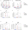
Characterization of CARCIK-CD19 cells. (A) PBMCs were transfected with the CAR-MNZ and CAR-BG2 plasmids and expanded to CIK for 21 days. Total cell number at the end of the culture is shown, starting from the same number of cells in both cases (10 × 106 cells). (B,C) Percentage (B) and MFI (C) of CAR expression on CD3+ cells at the end of culture. (D,E) Immunophenotype was analyzed at the end of the cultures (day 21) by flow cytometry, including percentages of CD3+, CD3+CD4+, CD3+CD8+, CD3+CD56+ (D) and effector memory populations (E); (FH) CAR+ cells were purified at day 10–14 by immunoselection. Percentages of CAR+ cells pre- and post-purification are shown. Representative flow cytometry histograms of CAR expression of non-purified and purified CARCIK-CD19 (respectively, CAR-MNZ 26.6%, 4588 MFI, and 97.1%, 4031 MFI; CAR-BG2 20.4%, 2641 MFI, and 96.8%, 3208 MFI). The results are the means and standard deviations of four to five experiments using different donors as starting material. (*: p < 0.05, ns: not significant).
Figure 3
Figure 3
In vitro activity. (A) Comparison of the killing activity in vitro using the calcein release assay. CIK (blue bars), CIK in presence of 10 ng/mL blinatumomab (red bars), CARCIK-MNZ (pink bars), and CARCIK-BG2 (green bars) at the end of culture were used as cytotoxic effector cells against the REH cell line at a 10:1, 3:1, and 1:1 E:T ratio. Percentage target lysis is shown as mean and standard deviation of six experiments. (B) Comparison of the proliferation ability in vitro (proliferation index) measured by flow cytometry on CFSE-stained effector cells, after co-culture with the target cell line REH, at a 10:1, 1:1, and 1:10 E:T ratio, for 4 days. Proliferation indexes are shown as mean and standard deviation of four experiments. (C,D) IFN-γ and IL-2 production was determined by intracytoplasmic flow cytometry after 6 h co-culture at 1:1 E:T ratio with REH cell line. Percentages of positive cells are shown as mean and standard deviation of five experiments. (E,F) The NFAT and NF-kB upon stimulation was measured by co-transfection of Lucia luciferase reporter plasmids into HuT 78 cell line wild type alone (as CIK, blue bars), or stimulated with 10 ng/mL blinatumomab (red bars), or stably transfected with CAR-MNZ (pink bars) or with CAR-BG2 (green bars) and co-cultured with REH cell line for 24 h. Relative luminescence units (RLUs) are shown as mean and standard deviation of five experiments. Columns represent the mean and bars the standard deviation (* p < 0.05, ** p < 0.01, *** p < 0.001).
Figure 4
Figure 4
Immunological synapse. Synapses events. All conditions are significatively different from each other (p < 0.01). Results are mean ± SD of three independent experiments performed.
Figure 5
Figure 5
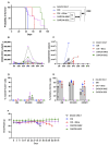
In vivo activity. (A) Kaplan–Meier survival curves of NSG mice engrafted with Daudi cells, left untreated (DAUDI only), or treated with CIKs, alone or receiving blinatumomab for the first three weeks, CARCIK-MNZ and CARCIK-BG2. (B) Analysis of hCD45+CD3+ cells/mL in the PB of animals treated with the different effectors. The data with DAUDI only condition (<63 hCD45+CD19+/mL) are not inserted in the graph for major clarity. (C) Analysis of hCD45+CD19+ cells/mL in the PB of animals treated with the different effectors or left untreated. (D) Percentages of hCD45+CD3+ cells in the BM, spleen, and kidney. (E) Percentages of hCD45+CD19+ cells in the BM, spleen, and kidney. (F) Mice body weight after the treatment. (** p < 0.01, *** p < 0.001, ns: not significant).

References

    1. Pievani A., Borleri G., Pende D., Moretta L., Rambaldi A., Golay J., Introna M. Dual-Functional Capability of CD3 + CD56 + CIK Cells, a T-Cell Subset That Acquires NK Function and Retains TCR-Mediated Specific Cytotoxicity. Blood. 2011;118:3301–3310. doi: 10.1182/blood-2011-02-336321. - DOI - PubMed
    1. Pievani A., Belussi C., Klein C., Rambaldi A., Golay J., Introna M. Enhanced Killing of Human B-Cell Lymphoma Targets by Combined Use of Cytokine-Induced Killer Cell (CIK) Cultures and Anti-CD20 Antibodies. Blood. 2011;117:510–518. doi: 10.1182/blood-2010-06-290858. - DOI - PubMed
    1. Franceschetti M., Pievani A., Borleri G., Vago L., Fleischhauer K., Golay J., Introna M. Cytokine-Induced Killer Cells Are Terminallydifferentiated Activated CD8 Cytotoxic T-EMRA Lymphocytes. Exp. Hematol. 2009;37:616–628.e2. doi: 10.1016/j.exphem.2009.01.010. - DOI - PubMed
    1. Introna M. CIK as Therapeutic Agents against Tumors. J. Autoimmun. 2017;85:32–44. doi: 10.1016/j.jaut.2017.06.008. - DOI - PubMed
    1. Valgardsdottir R., Capitanio C., Texido G., Pende D., Cantoni C., Pesenti E., Rambaldi A., Golay J., Introna M. Direct Involvement of CD56 in Cytokine-Induced Killer-Mediated Lysis of CD56+ Hematopoietic Target Cells. Exp. Hematol. 2014;42:1013–1021.e1. doi: 10.1016/j.exphem.2014.08.005. - DOI - PubMed

LinkOut - more resources