

Radiomics to predict PNI in ESCC
- PMID: 39311949
- PMCID: PMC11947035
- DOI: 10.1007/s00261-024-04562-8
Radiomics to predict PNI in ESCC
Abstract
Objective: This study aimed to investigate whether contrast-enhanced computed tomography (CECT) based radiomics analysis could noninvasively predict the perineural invasion (PNI) in esophageal squamous cell carcinoma (ESCC).
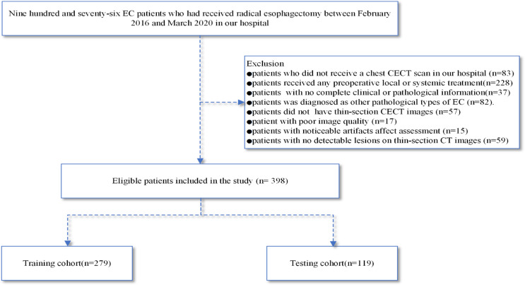
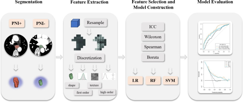
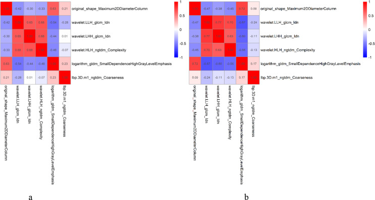
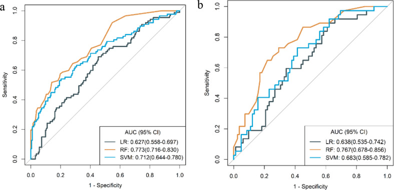
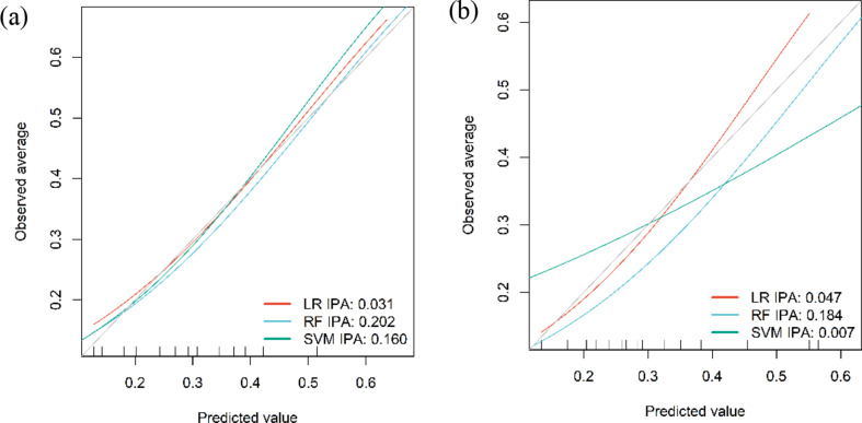
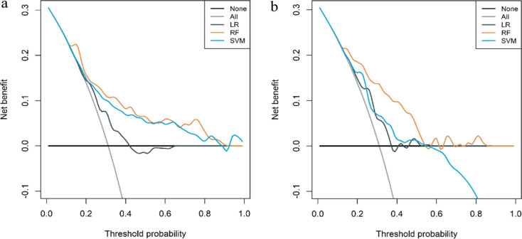
Methods: 398 patients with ESCC who underwent resection between February 2016 and March 2020 were retrospectively enrolled in this study. Patients were randomly divided into training and testing cohorts in a 7:3 ratio. Radiomics analysis was performed on the arterial phase images of CECT scans. From these images, 1595 radiomics features were initially extracted. The intraclass correlation coefficient (ICC), wilcoxon rank-sum test, spearman correlation analysis, and boruta algorithm were used for feature selection. Logistic regression (LR), random forest (RF), and support vector machine (SVM) models were established to preidict the PNI status. The performance of these radiomics models was assessed by the area under the receiver operating characteristic curve (AUC). Decision curve analysis (DCA) was conducted to evaluate their clinical utility.
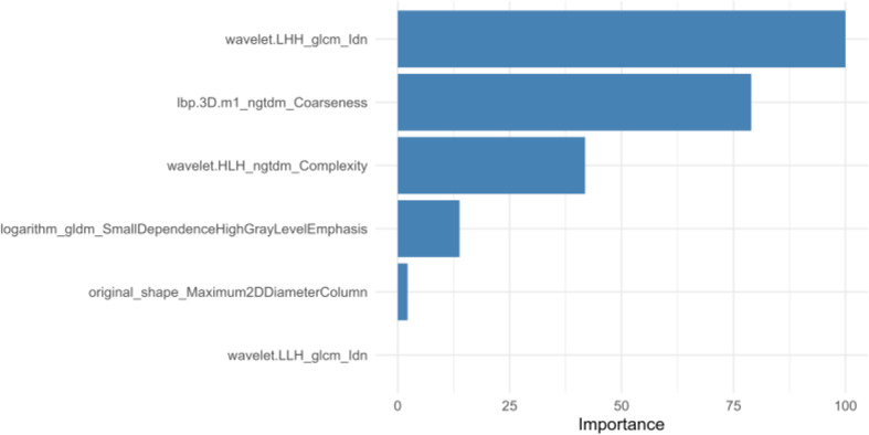
Results: Six radiomics features were retained to build the radiomics models. Among these models, the random forest (RF) model demonstrated superior performance. In the training cohort, the AUC value of the RF model was 0.773, compared to 0.627 for the logistic regression (LR) model and 0.712 for the support vector machine (SVM) model. Similarly, in the testing cohort, the RF model achieved an AUC value of 0.767, outperforming the LR model at 0.638 and the SVM model at 0.683. Decision curve analysis (DCA) suggested that the RF radiomics model exhibited the highest clinical utility.
Conclusions: CECT-based radiomics analysis, particularly utilizing the RF, can noninvasively predict the PNI in ESCC preoperatively. This novel approach could enhance patient management by providing personalized information, thereby facilitating the development of individualized treatment strategies for ESCC patients.
Keywords: Computed tomography; Contrast-enhanced; Esophageal squamous cell carcinoma; Perineural invasion; Radiomics.
© 2024. The Author(s).
Conflict of interest statement
Declarations. Competing interests: The authors declare no competing interests.
Figures
Similar articles
-
Prediction of lymphovascular invasion in esophageal squamous cell carcinoma by computed tomography-based radiomics analysis: 2D or 3D ?Cancer Imaging. 2024 Oct 17;24(1):141. doi: 10.1186/s40644-024-00786-5. Cancer Imaging. 2024. PMID: 39420415 Free PMC article.
-
Computed tomography-based radiomics nomogram for prediction of lympho-vascular and perineural invasion in esophageal squamous cell cancer patients: a retrospective cohort study.Cancer Imaging. 2024 Oct 4;24(1):131. doi: 10.1186/s40644-024-00781-w. Cancer Imaging. 2024. PMID: 39367492 Free PMC article.
-
CT-based radiomics model for predicting perineural invasion status in gastric cancer.Abdom Radiol (NY). 2025 May;50(5):1916-1926. doi: 10.1007/s00261-024-04673-2. Epub 2024 Nov 6. Abdom Radiol (NY). 2025. PMID: 39503776
-
CT Multidimensional Radiomics Combined with Inflammatory Immune Score For Preoperative Prediction of Pathological Grade in Esophageal Squamous Cell Carcinoma.Acad Radiol. 2025 May;32(5):2667-2678. doi: 10.1016/j.acra.2024.12.030. Epub 2025 Jan 13. Acad Radiol. 2025. PMID: 39809604
-
Computed tomography-based radiomics analysis to predict lymphovascular invasion in esophageal squamous cell carcinoma.Br J Radiol. 2022 Feb 1;95(1130):20210918. doi: 10.1259/bjr.20210918. Epub 2021 Dec 15. Br J Radiol. 2022. PMID: 34908477 Free PMC article.
References
-
- Uhlenhopp DJ, Then EO, Sunkara T, Gaduputi V (2020) Epidemiology of esophageal cancer: update in global trends, etiology and risk factors. Clin J Gastroenterol 13:1010–1021. doi:10.1007/s12328-020-01237-x - PubMed
-
- Siegel RL, Miller KD, Jemal A (2020) Cancer statistics, 2020. CA Cancer J Clin 70:7–30. doi:10.3322/caac.21590 - PubMed
-
- Liebig C, Ayala G, Wilks JA, Berger DH, Albo D (2009) Perineural invasion in cancer: a review of the literature. Cancer 115:3379–3391. doi:10.1002/cncr.24396 - PubMed
MeSH terms
Substances
LinkOut - more resources
Full Text Sources
Medical
