. 2024 Sep 23;14(1):149.
doi: 10.1186/s13613-024-01378-z.

Impact of airway closure and lung collapse on inhaled nitric oxide effect in acute lung injury: an experimental study

Affiliations

Impact of airway closure and lung collapse on inhaled nitric oxide effect in acute lung injury: an experimental study

Mariangela Pellegrini et al. Ann Intensive Care. .

Abstract

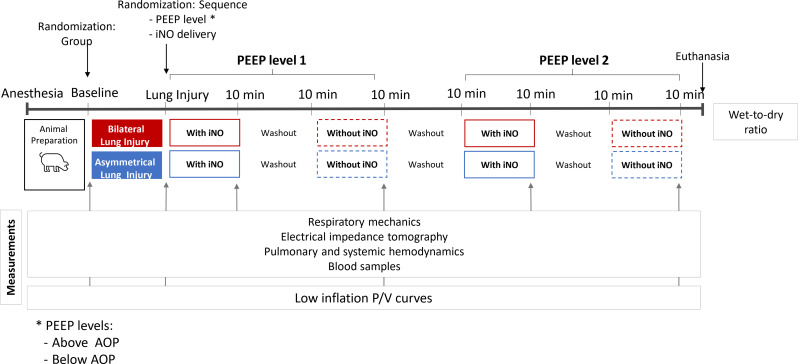
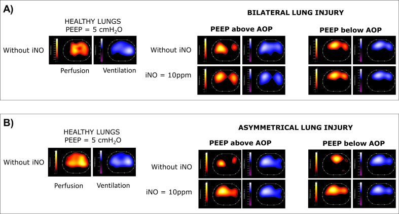
Background: Efficacy of inhaled therapy such as Nitric Oxide (iNO) during mechanical ventilation may depend on airway patency. We hypothesized that airway closure and lung collapse, countered by positive end-expiratory pressure (PEEP), influence iNO efficacy. This could support the role of an adequate PEEP titration for inhalation therapy. The main aim of this study was to assess the effect of iNO with PEEP set above or below the airway opening pressure (AOP) generated by airway closure, on hemodynamics and gas exchange in swine models of acute respiratory distress syndrome. Fourteen pigs randomly underwent either bilateral or asymmetrical two-hit model of lung injury. Airway closure and lung collapse were measured with electrical impedance tomography as well as ventilation/perfusion ratio (V/Q). After AOP detection, the effect of iNO (10ppm) was studied with PEEP set randomly above or below regional AOP. Respiratory mechanics, hemodynamics, and gas-exchange were recorded.

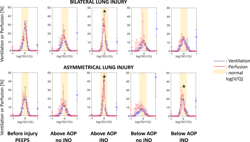
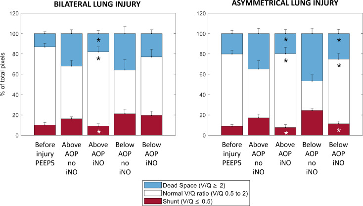
Results: All pigs presented airway closure (AOP > 0.5cmH2O) after injury. In bilateral injury, iNO was associated with an improved mean pulmonary pressure from 49 ± 8 to 42 ± 7mmHg; (p = 0.003), and ventilation/perfusion matching, caused by a reduction in pixels with low V/Q and shunt from 16%[IQR:13-19] to 9%[IQR:4-12] (p = 0.03) only at PEEP set above AOP. iNO had no effect on hemodynamics or gas exchange for PEEP below AOP (low V/Q 25%[IQR:16-30] to 23%[IQR:14-27]; p = 0.68). In asymmetrical injury, iNO improved pulmonary hemodynamics and ventilation/perfusion matching independently from the PEEP set. iNO was associated with improved oxygenation in all cases.

Conclusions: In an animal model of bilateral lung injury, PEEP level relative to AOP markedly influences iNO efficacy on pulmonary hemodynamics and ventilation/perfusion match, independently of oxygenation.

Keywords: (3 to 10) nitric oxide; Acute respiratory distress syndrome; Airway closure; Mechanical ventilation.


Conflict of interest statement

All authors disclose any financial and personal conflicts of interest.

Figures

Fig. 1
Fig. 1
Study protocol overview. Abbreviations: PEEP, Positive End-Expiratory Pressure; iNO, inhaled nitric oxide; aop, airway opening pressure. *: PEEP levels above/below AOP; **: this flowchart represents a sequence of 10-min with iNO at 10 ppm followed by 10-min without iNO
Fig. 2
Fig. 2
Electrical impedance tomography in a representative example of bilateral (A) and asymmetrical (B) lung injury. For each studied condition (i.e., healthy lung - HL, injured lung at PEEP above/below AOP, with/without iNO two derived maps are reported: 1) the perfusion map reported in red-orange-yellow scale (where yellow represents high perfusion and red low perfusion); 2) the ventilation map reported in blue-sky blue-white (where white corresponds to highly ventilated areas). Abbreviations: PEEP: positive end expiratory pressure; iNO: inhaled nitric oxide; AOP: airway opening pressure
Fig. 3
Fig. 3
Highest global airway opening pressure (AOP) by group
Fig. 4
Fig. 4
Effects of iNO on pulmonary hemodynamics. (A) Mean Pulmonary Artery Pressure (mPAP) and (B) Transpulmonary Gradient Pressure (TPG) response to inhaled nitric oxide according to Positive End-Expiratory Pressure (PEEP), i.e., below Airway Opening Pressure (AOP) or above AOP, and type of lung injury, i.e., bilateral or asymmetrical. Note: p-values are from univariate analysis (paired t-test or Wilcoxon test)
Fig. 5
Fig. 5
Ventilation/perfusion analysis based on electrical impedance tomography. x-axis: log10(V/Q) where 0 indicates ventilation equal to perfusion; -1 indicates perfusion 10 times higher than ventilation, and + 1 indicates ventilation 10 times higher than perfusion. Moreover, the variable on the x-axis is analysed and reported as discrete, where all original values are approximated to the closest fist decimal number between − 1 and + 1, originating 21 possible values. Log(V/Q) = -1 reports all log(V/Q) values equal or lower than − 1. Log(V/Q) = 1 reports all log(V/Q) values equal or higher than + 1. y-axis: regional distribution of the fraction of ventilation (blue) and perfusion (red), indicated as percentage of the total impedance in the corresponding (ventilation or perfusion) map. Values reported as median (IQR) and represented as by open circles and whiskers. The solid lines correspond to the best fitting curves (with 95%CI). *: to mark significant differences compare to the correspondig ventilatory condition without iNO (Two-sided Wilcoxon signed rank test; α = 0.05). The yellow area indicating normal V/Q between 0.5 and 2 corresponding to log(V/Q) between − 0.3 and 0.3. Abbreviations: V/Q: ventilation/perfusion ratio; PEEP: positive end-expiratory pressure; AOP: airway opening pressure; iNO: inhaled nitric oxide
Fig. 6
Fig. 6
Percentages of pixels for each lung compartment: (1) shunt (red) defined as log(V/Q) lower than or equal to -0.3 or as V/Q lower than or equal to 0.5; (2) normal V/Q (white) defined as a V/Q between 0.5 and 2; (3) dead space (blue) defined as log(V/Q) equal to or higher than 0.3 or as V/Q equal to or higher than 2. Values reported as mean + SEM. Abbreviations: V/Q: ventilation/perfusion ratio; PEEP: positive end-expirtory pressure; AOP: airway opening pressure; iNO: inhaled nitric oxide. ). *: to mark significant differences compare to the correspondig ventilatory condition without iNO (Two-sided Wilcoxon signed rank test; α = 0.05)

References

    1. The ARDS Definition Task Force. Acute respiratory distress syndrome: the Berlin definition. JAMA. 2012;307:2526–33. - PubMed
    1. Bellani G, Laffey JG, Pham T, Fan E, Brochard L, Esteban A, et al. Epidemiology, patterns of Care, and mortality for patients with Acute Respiratory Distress Syndrome in Intensive Care Units in 50 countries. JAMA. 2016;315:788–800. - PubMed
    1. Katzenstein AL, Bloor CM, Leibow AA. Diffuse alveolar damage–the role of oxygen, shock, and related factors. A review. Am J Pathol. 1976;85:209–28. - PMC - PubMed
    1. Thille AW, Esteban A, Fernández-Segoviano P, Rodriguez J-M, Aramburu J-A, Vargas-Errázuriz P, et al. Chronology of histological lesions in acute respiratory distress syndrome with diffuse alveolar damage: a prospective cohort study of clinical autopsies. Lancet Respir Med. 2013;1:395–401. - PubMed
    1. Zapol WM, Jones R. Vascular components of ARDS: clinical pulmonary hemodynamics and morphology. Am Rev Respir Dis. 1987;136:471–4. - PubMed

LinkOut - more resources