. 2024 Dec;49(12):3297-3307.
doi: 10.1007/s11064-024-04230-9. Epub 2024 Sep 23.

Zona Incerta GABAergic Neurons Facilitate Emergence from Isoflurane Anesthesia in Mice

Affiliations

Zona Incerta GABAergic Neurons Facilitate Emergence from Isoflurane Anesthesia in Mice

Hong Chen et al. Neurochem Res. 2024 Dec.

Abstract

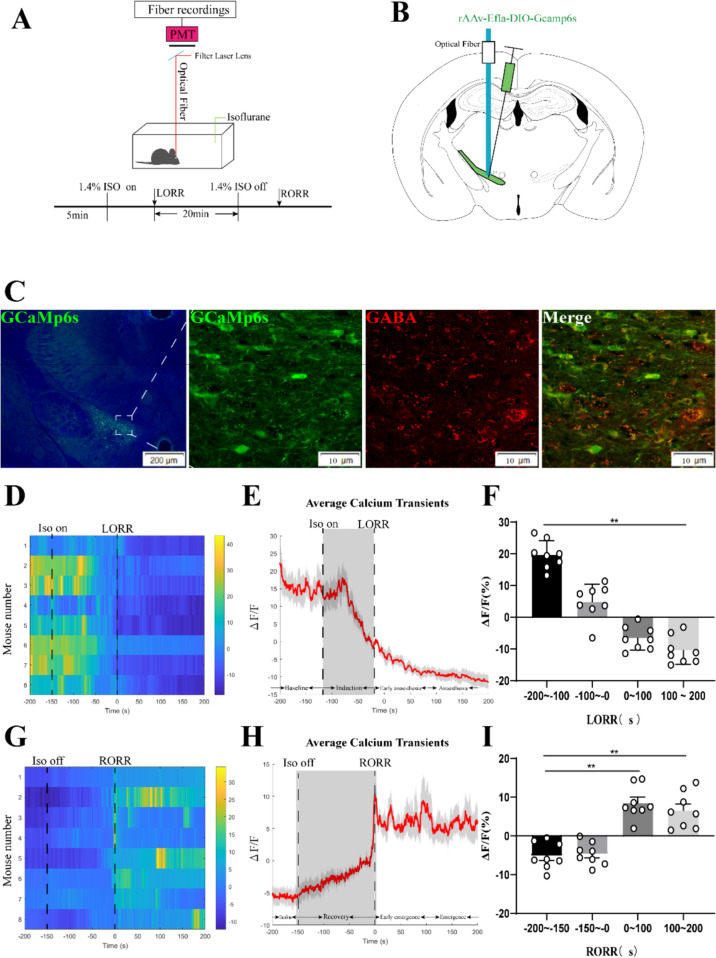
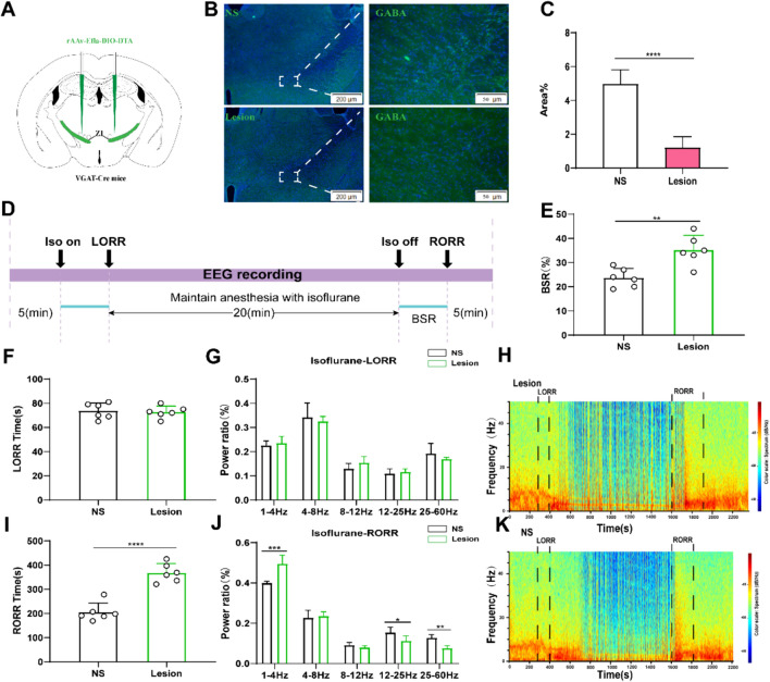
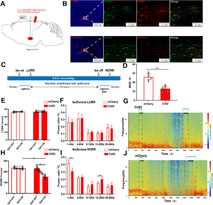
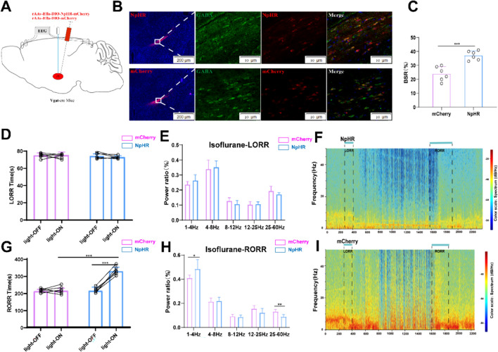
The zona incerta (ZI) predominantly consists of gamma-aminobutyric acid (GABAergic) neurons, located adjacent to the lateral hypothalamus. GABA, acting on GABAA receptors, serves as a crucial neuromodulator in the initiation and maintenance of general anesthesia. In this study, we aimed to investigate the involvement of ZI GABAergic neurons in the general anesthesia process. Utilizing in-vivo calcium signal optical fiber recording, we observed a decrease in the activity of ZI GABAergic neurons during isoflurane anesthesia, followed by a significant increase during the recovery phase. Subsequently, we selectively ablated ZI GABAergic neurons to explore their role in general anesthesia, revealing no impact on the induction of isoflurane anesthesia but a prolonged recovery time, accompanied by a reduction in delta-band power in mice under isoflurane anesthesia. Finally, through optogenetic activation/inhibition of ZI GABAergic neurons during isoflurane anesthesia, we discovered that activation of these neurons facilitated emergence without affecting the induction process, while inhibition delayed emergence, leading to fluctuations in delta band activity. In summary, these findings highlight the involvement of ZI GABAergic neurons in modulating the emergence of isoflurane anesthesia.

Keywords: Emergence time; GABAergic Neurons; Isoflurane Anesthesia; Optogenetic; Zona Incerta.


Conflict of interest statement

The authors declare no competing interests.

Figures

Fig. 1
Fig. 1
Phase-dependent calcium alterations in ZI GABAergic neurons during isoflurane anaesthesia. Top: Schematic diagram of fiber photometry recording during isoflurane anesthesia in freely moving mice. Bottom: Timeline for quantifying the LORR and RORR of isoflurane anesthesia. (PMT, Photomultiplier tube is a kind of detection device that can convert weak light signals into electronic signals and amplify them). B Schematic of calcium signal recording model into the ZI of a VGAT -Cre mouse. C Expression of GCaMP6f in the ZI of a VGAT -Cre mouse. Viral expression (GCaMP6s, green) in the ZI and colabeling with GABA neurons (GABA immunofluorescence, red). D Individual transitions from wakefulness to isoflurane-induced LORR with color-coded fluorescent intensities (LORR were represented the point of 0). E Average responses from the state transitions during the process of induction expressed as the mean (red) ± SEM (shaded). (F) The fluorescence calcium signals significantly reduced after isoflurane-induced unconsciousness (The baseline (wake: − 200 to − 100 s) vs. anaesthesia period (100 to 200 s), P = 0.0475; n = 8), the paired Student’s t-tests). G Individual transitions from isoflurane anesthesia state to arousal with color-coded fluorescent intensities (RORR were represented the point of 0). H Average responses from the state transitions during the process of recovery expressed as the mean (red) ± SEM (shaded). I The fluorescence calcium signals ascended after RORR (The baseline (anaesthesia: − 200 to − 150 s) vs. early emergence period (0 to 100 s); P = 0.0055; The baseline: (anaesthesia: − 200 to − 150s) vs. emergence period (100 to 200 s), P = 0.0026; the paired Student’s t-tests; n = 8, *P < 0.05, **P < 0.01)
Fig. 2
Fig. 2
Lesion of ZI GABAergic neurons delayed arousal from isoflurane anesthesia. A Schematic showing bilateral injection of Cre-independent AAVs into the ZI. Image showing GABA staining from a mouse with specific ZI lesion using AAV-CAG-DIO-DTA (NS represents normal saline). C Proportions of GABA-fluorescent area in ZI, indicating that the lesion group animals were selectively ablated ZI GABAergic neurons (P = 0.0001 by independent-samples t-test; n = 6 per group). D Protocol for behavioral and electroencephalogram (EEG) recording of induction time and emergence time. Spectrograms of EEG power during the isoflurane anaesthesia period in the control group. E BSR at recovery process of isoflurane in control group and lesion group (P = 0.0001 by independent-samples t-test; n = 6 per group). F The induction time in control group and lesion group during the maintenance of 1.4% isoflurane anesthesia. G Lesion of ZI GABAergic neurons displaying no significant change in the power ratios of the EEG. Spectrograms of EEG power during the isoflurane anaesthesia period in lesion group (H) and the control group (K). I Lesion of ZI GABAergic neurons peolonged the recovery time from 1.4% isoflurane anesthesia. (J) During the recovery time, lesion of ZI GABAergic neurons significantly altered the power ratios of the EEG. (δ band: lesion group vs. control group; P = 0.0003 by independent-samples t-test;β band: lesion group vs. control group; P = 0.018 by independent-samples t-test, γ band: lesion group vs. control group; P = 0.01 by independent-samples t-test; n = 6 per group)
Fig. 3
Fig. 3
Optogenetic activation of ZI GABAergic neurons facilitates arousal from isoflurane anesthesia. A Diagram of the optogenetic virus injection and stimulation sites in ZI. B Expression of virus (ChR2 and mCherry, red) in ZI GABAergic neurons and colabeling with GABA (GABA immunofluorescence, green). C Protocol for optogenetic activation during isoflurane anesthesia. D Optical activation of ZI GABAergic neurons reduced the ratio of BSR in the recovery period. E Optical activation of ZI GABAergic neurons did not change induction time (LORR: mcherry-light-on vs. ChR2-light-on, P = 0.6, independent-simples t-test; ChR2-light-on vs. ChR2-light-off. P = 0.2, paired t-test). F, I Comparison of each EEG frequency band between the two groups during optogenetic activation of ZI GABAergic neurons. Spectrograms of EEG power during the isoflurane anaesthesia period in ChR2 group G and the mCherry group (J). H Optical activation of ZI GABAergic neurons shortened emergence time from 1.4% isoflurane anesthesia (RORR: mcherry-light-on vs. ChR2-light-on, P = 0.0009, independent-simples t-test; ChR2-light-on vs. ChR2-light-off. P = 0.0002, paired t-test). Representative EEG heatmap of the process in the two groups
Fig. 4
Fig. 4
Optogenetic inhibition of ZI GABAergic neurons slow down recovery from isoflurane anesthesia. A Schematic of optogenetic inhibition of NpHR-expressing ZI GABAergic neurons with EEG recordings B Expression of NpHR (red) and GABA immunofluorescence (green) in the ZI. C Optical inhibition of ZI GABAergic neurons reduced the ratio of BSR in the recovery period. D Optical stimulation of ZI GABAergic neurons did not alter the induction time of isoflurane anesthesia. E Optical inhibition of ZI GABAergic neurons did not change the band distribution of EEG power during the induction time. F Representative EEG heatmap of NpHR group. G Optogenetic inhibition of ZI GABAergic neurons delayed the emergence from isoflurane anesthesia. H Optogenetic inhibition of ZI GABAergic neurons regulated the band distribution of EEG power during RORR processes of isoflurane anesthesia. Representative EEG heatmap of control group

Similar articles

References

    1. Franks NP, Wisden W (2021) The inescapable drive to sleep: overlapping mechanisms of sleep and sedation. Science 374(6567):556–559 - PubMed
    1. Liu C et al (2021) Lateral habenula glutamatergic neurons modulate isoflurane anesthesia in mice. Front Mol Neurosci 14:628996 - PMC - PubMed
    1. Wang D et al (2021) GABAergic neurons in the dorsal-intermediate lateral septum regulate sleep-wakefulness and anesthesia in mice. Anesthesiology 135(3):463–481 - PubMed
    1. Franks NP (2008) General anaesthesia: from molecular targets to neuronal pathways of sleep and arousal. Nat Rev Neurosci 9(5):370–386 - PubMed
    1. Vlasov K et al (2021) Activation of GABAergic neurons in the Rostromedial Tegmental Nucleus and other Brainstem regions promotes sedation and facilitates sevoflurane anesthesia in mice. Anesth Analg 132(4):e50–e55 - PMC - PubMed

LinkOut - more resources