. 2024 Oct 10;67(19):17087-17100.
doi: 10.1021/acs.jmedchem.4c00292. Epub 2024 Sep 23.

Use of the Novel Site-Directed Enzyme Enhancement Therapy (SEE-Tx) Drug Discovery Platform to Identify Pharmacological Chaperones for Glutaric Acidemia Type 1

Affiliations

Use of the Novel Site-Directed Enzyme Enhancement Therapy (SEE-Tx) Drug Discovery Platform to Identify Pharmacological Chaperones for Glutaric Acidemia Type 1

Madalena Barroso et al. J Med Chem. .

Abstract

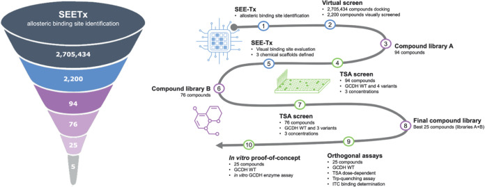
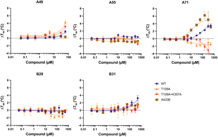
Allosteric regulators acting as pharmacological chaperones hold promise for innovative therapeutics since they target noncatalytic sites and stabilize the folded protein without competing with the natural substrate, resulting in a net gain of function. Exogenous allosteric regulators are typically more selective than active site inhibitors and can be more potent than competitive inhibitors when the natural substrate levels are high. To identify novel structure-targeted allosteric regulators (STARs) that bind to and stabilize the mitochondrial enzyme glutaryl-CoA dehydrogenase (GCDH), the computational site-directed enzyme enhancement therapy (SEE-Tx) technology was applied. SEE-Tx is an innovative drug discovery platform with the potential to identify drugs for treating protein misfolding disorders, such as glutaric acidemia type 1 (GA1) disease. Putative allosteric regulators were discovered using structure- and ligand-based virtual screening methods and validated using orthogonal biophysical and biochemical assays. The computational approach presented here could be used to discover allosteric regulators of other protein misfolding disorders.


Conflict of interest statement

The authors declare the following competing financial interest(s): EC, AD, AMG, MM, and XB completed the research and the authorship of this article within the scope of their employ-ment. EC, AD, AMG, and XB are employees of Gain Therapeu-tics Sucursal en Espaa and MM is an employee and sharehold-er of Minoryx Therapeutics. ACM and SWG are co-founders and shareholders of iniuva GmbH.

Figures

Figure 1
Figure 1
Putative allosteric binding site of glutaryl-CoA dehydrogenase (GCDH) monomer identified using the SEE-Tx platform. (A) Overview of GCDH structure (1SIR.pdb) showing the allosteric binding site (green area) at the interface of the α-helical bundle amino-terminal domain (light orange) and the α-helical domain at the carboxyl terminus (light blue). The active site is located at the opposite side of the protein, at a distance of 12 Å. S-4-nitrobutyryl-CoA (an alternative substrate) and FAD (cofactor) are shown as cyan and yellow sticks, respectively. (B) MDmix identified binding hot spots at the allosteric site corresponding to polar (red) and lipophilic (green) interactions. (C) Predicted binding mode of compound A19 within the allosteric pocket. H-bonding interactions are indicated by yellow dashed lines.
Figure 2
Figure 2
Drug discovery platform applied for the identification of potential pharmacological chaperones for glutaric acidemia type 1. SEE-Tx methodology uses a structure-based approach to identify putative binders for glutaryl-CoA dehydrogenase (GCDH). This was followed by a step-by-step validation approach using thermal shift assays and additional orthogonal biophysical and biochemical assays, which allowed the identification of 5 validated allosteric regulators of GCDH.
Figure 3
Figure 3
Dose-dependent effect on thermal stability of glutaryl-CoA dehydrogenase (GCDH) wild type (WT) in the presence of (A) compounds A19, A34, A35, A41, A49, A58 and A71 from library A, and (B) compounds B12, B14, B15, B29, B30, B34, B47, B49, and B56 from library B, at three different concentrations (10, 30, and 100 μM). Most compounds have a shift in Tm (melting temperature: the temperature at which 50% of a protein is unfolded) relative to the baseline (“No compound,” dotted line), suggesting protein stabilization. Tm for each compound concentration is shown (circle, triangle, square, for 10, 30, and 100 μM, respectively) ± standard deviation (transparent bar behind Tm value). Gray shaded blocks represent ΔTm shifts of >1 °C, >2 °C and >3 °C.
Figure 4
Figure 4
Tryptophan fluorescence quenching for glutaryl-CoA dehydrogenase (GCDH) wild type (WT) with increasing compound concentrations of the 25 selected compounds, comprising two main scaffolds and various singletons (“Others 1” and “Others 2”). Intrinsic fluorescence of GCDH WT is quenched due to the binding of compounds at increasing concentrations. F0, initial fluorescence intensity; F, fluorescence intensity in the presence of a quenching agent.
Figure 5
Figure 5
Isothermal titration calorimetry (ITC) analysis of the interaction of glutaryl-CoA dehydrogenase (GCDH) wild type (WT) with compounds A41, A49, A58, A66 and A71. Crotonyl-CoA, the GCDH enzyme reaction product, was used as a positive binding control. ΔH (change in enthalpy derived from integration of the heat peak intensities) is plotted against the GCDH WT/compound molar ratio (based on monomer concentration).
Figure 6
Figure 6
Effect of the final compound library on the catalytic activity of recombinant GCDH WT. The selected scaffolds are color-coded (scaffold 1 in blue; scaffold 2 in green; other individual scaffolds in gray). The mean values and standard deviations were determined from three independent assays (*, p < 0.05; **, p < 0.005; ***, p < 0.0005). DMSO, dimethyl sulfoxide; GCDH, glutaryl-CoA dehydrogenase; WT, wild type.
Figure 7
Figure 7
Effect of the 5 leading compounds on enzymatic activity of recombinant GCDH WT and variants R88C and V400M. Enzyme kinetics were measured in the presence of different compound concentrations (0.25–125 μM). Enzyme activity is shown as mean UI/nmol of protein (±standard deviation). Two experimental replicates were used. WT, wild type.
Figure 8
Figure 8
Competition assay for proteins GCDH WT with compound A49 (left, purple) and GCDH V400M and A71 (right, green). Enzyme kinetics were measured in the presence of different substrate concentrations (0–1.5 mM). Enzyme activity is shown as mean UI/nmol of protein (±standard deviation). A Michaelis–Menten equation was used to fit the data (line). Two experimental replicates were used. GCDH, glutaryl-CoA dehydrogenase; WT, wild type.
Figure 9
Figure 9
Dose-dependent effect on thermal stability of glutaryl-CoA dehydrogenase (GCDH) wild type (WT) and mutations mapping to the proposed allosteric binding site in the presence of lead compounds A49, A55, A71, B29, and B31. Thermal shift assays were performed for each protein variant (WT, A433E, and allosteric pocket mutant T109A, and double mutant T109A and K357A), using different compound concentrations from 0.05 to 500 μM. Variant A433E was used as an extra control for a pocket-independent mutation. A Tm (melting temperature: the temperature at which 50% of a protein is unfolded) was calculated for each concentration, and the thermal shifts (ΔTms, difference of Tm in the presence and absence of compound) were plotted against compound concentration. ΔTms are shown as mean and error bars corresponding to standard deviation (n = 3).

Similar articles

Cited by

References

    1. Boy N.; Mengler K.; Heringer-Seifert J.; Hoffmann G. F.; Garbade S. F.; Kölker S. Impact of Newborn Screening and Quality of Therapy on the Neurological Outcome in Glutaric Aciduria Type 1: A Meta-Analysis. Genet. Med. 2021, 23 (1), 13–21. 10.1038/s41436-020-00971-4. - DOI - PMC - PubMed
    1. Kölker S.; Garbade S. F.; Greenberg C. R.; Leonard J. V.; Saudubray J. M.; Ribes A.; Kalkanoglu H. S.; Lund A. M.; Merinero B.; Wajner M.; et al. Natural History, Outcome, and Treatment Efficacy in Children and Adults with Glutaryl-CoA Dehydrogenase Deficiency. Pediatr. Res. 2006, 59 (6), 840–847. 10.1203/01.pdr.0000219387.79887.86. - DOI - PubMed
    1. Boy N.; Mühlhausen C.; Maier E. M.; Heringer J.; Assmann B.; Burgard P.; Dixon M.; Fleissner S.; Greenberg C. R.; Harting I.; et al. Proposed Recommendations for Diagnosing and Managing Individuals with Glutaric Aciduria Type I: Second Revision. J. Inherit. Metab. Dis. 2017, 40 (1), 75–101. 10.1007/s10545-016-9999-9. - DOI - PubMed
    1. Strauss K. A.; Williams K. B.; Carson V. J.; Poskitt L.; Bowser L. E.; Young M.; Robinson D. L.; Hendrickson C.; Beiler K.; Taylor C. M.; et al. Glutaric Acidemia Type 1: Treatment and Outcome of 168 Patients over Three Decades. Mol. Genet. Metab. 2020, 131 (3), 325–340. 10.1016/j.ymgme.2020.09.007. - DOI - PubMed
    1. Märtner E. M. C.; Thimm E.; Guder P.; Schiergens K. A.; Rutsch F.; Roloff S.; Marquardt I.; Das A. M.; Freisinger P.; Grünert S. C.; et al. The Biochemical Subtype is a Predictor for Cognitive Function in Glutaric Aciduria Type 1: A National Prospective Follow-Up Study. Sci. Rep. 2021, 11 (1), 1930010.1038/s41598-021-98809-9. - DOI - PMC - PubMed

MeSH terms

Substances

Supplementary concepts