. 2024 Sep 23;19(9):e0308648.
doi: 10.1371/journal.pone.0308648. eCollection 2024.

Matrix metalloproteinases mediate influenza A-associated shedding of the alveolar epithelial glycocalyx

Affiliations

Matrix metalloproteinases mediate influenza A-associated shedding of the alveolar epithelial glycocalyx

Kaitlyn R Schaaf et al. PLoS One. .

Abstract

Background: The alveolar epithelium is protected by a heparan sulfate-rich, glycosaminoglycan layer called the epithelial glycocalyx. It is cleaved in patients with acute respiratory distress syndrome (ARDS) and in murine models of influenza A (IAV) infection, shedding fragments into the airspace from the cell surface. Glycocalyx shedding results in increased permeability of the alveolar-capillary barrier, amplifying acute lung injury. The mechanisms underlying alveolar epithelial glycocalyx shedding in IAV infection are unknown. We hypothesized that induction of host sheddases such as matrix metalloproteinases (MMPs) during IAV infection results in glycocalyx shedding and increased lung injury.

Materials and methods: We measured glycocalyx shedding and lung injury during IAV infection with and without treatment with the pan-MMP inhibitor Ilomastat (ILO) and in an MMP-7 knock out (MMP-7KO) mouse. C57BL/6 or MMP-7KO male and female mice were given IAV A/PR/8/34 (H1N1) at 30,000 PFU/mouse or PBS intratracheally. For some experiments, C56BL/6 mice were infected in the presence of ILO (100mg/kg) or vehicle given daily by IP injection. Bronchoalveolar lavage (BAL) and lung tissue were collected on day 1, 3, and 7 for analysis of glycocalyx shedding (BAL Syndecan-1) and lung injury (histology, BAL protein, BAL cytokines, BAL immune cell infiltrates, BAL RAGE). Expression and localization of the sheddase MMP-7 and its inhibitor TIMP-1 was examined by RNAScope. For in vitro experiments, MLE-12 mouse lung epithelial cells were cultured and treated with active or heat-inactivated heparinase (2.5 U/mL) prior to infection with IAV (MOI 1) and viral load and MMP-7 and TIMP-1 expression analyzed.

Results: IAV infection caused shedding of the epithelial glycocalyx into the BAL. Inhibition of MMPs with ILO reduced glycocalyx shedding by 36% (p = 0.0051) and reduced lung epithelial injury by 40% (p = 0.0404). ILO also reduced viral load by 68% (p = 0.027), despite having no significant effect on lung cytokine production. Both MMP-7 and its inhibitor TIMP-1 were upregulated in IAV infected mice: MMP-7 colocalized with IAV, while TIMP-1 was limited to cells adjacent to infection. However, MMP-7KO mice had similar glycocalyx shedding, epithelial injury, and viral load compared to WT littermates, suggesting redundancy in MMP sheddase function in the lung. In vitro, heparinase treatment before infection led to a 52% increase in viral load (p = 0.0038) without altering MMP-7 or TIMP-1 protein levels.

Conclusions: Glycocalyx shedding and MMPs play key roles in IAV-induced epithelial injury, with significant impact on IAV viral load. Further studies are needed to understand which specific MMPs regulate lung epithelial glycocalyx shedding.


Conflict of interest statement

The authors have declared that no competing interests exist.

Figures
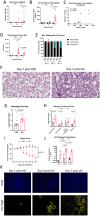
Fig 1
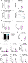
Fig 1. IAV causes alveolar glycocalyx shedding during acute infection in mice.
Wild-type C57BL/6 mice were intranasally administered PR8 influenza or PBS. On day 7, IAV infected mice had (A) increased shedding of the alveolar epithelial glycocalyx as measured by glycocalyx anchoring protein Syndecan-1 in bronchoalveolar lavage (BAL) fluid (n = 6–11 [7 male, 4 female]; overall p = 0.015 for infection, two-way ANOVA with significant post-hoc relationships within days indicated) and (B) increased alveolar-capillary barrier permeability as measured by total BAL protein (n = 10–14 [10 male, 4 female], overall p<0.0001 for infection, overall p<0.0001 for time, two-way ANOVA with significant post-hoc relationships within days indicated). (C) Glycocalyx shedding correlates with alveolar barrier permeability (n = 16 [12 male, 4 female], Spearman’s Correlation). IAV infected mice also have (D) increased total inflammatory cells in BAL (n = 10–15 [11 male, 4 female], overall p<0.0001 for infection, overall p<0.0001 for time, two-way ANOVA with significant post-hoc relationships within days indicated), with (E) increased neutrophil influx at day 3 and day 7 (P<0.001 for each, Mann-Whitney U for each day with Holm-Sidak correction). (F) Representative histologic images (hematoxylin and eosin) from control and IAV infected lungs at day 7 are shown and (G) lung injury quantification by assessment of ten non-overlapping visual fields using a validated lung injury score indicates significant lung injury in IAV mice (n = 6–10 [5 male, 5 female], Mann-Whitney U). (H) Components of the lung injury score show that inflammation, hemorrhage, and edema are the primary manifestations of IAV-induced lung injury (Mann-Whitney U for each category with Holm-Sidak correction). (I) IAV mice have increased weight loss beginning at day 3 (n = 9–42 [22 male, 20 female]; mixed model ANOVA). (J) IAV viral load peaks at day 3, as measured by relative expression (ΔΔCt) analysis to IAV day 1 of RT-qPCR of IAV genome segment 8 (n = 8–11 [9 male, 4 female], overall p = 0.001 for infection and overall p = 0.012 for time, two-way ANOVA with significant post-hoc relationships indicated). (K) IAV infection progresses from larger airways on day 1, to smaller airways on day 3, to alveoli on day 7. RNA in situ hybridization shows IAV genome segment 8 in yellow and DAPI nuclear counterstain in blue. There are no statistically significant sex-specific differences in these parameters.
Fig 2
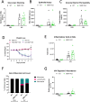
Fig 2. Pan-MMP inhibitor Ilomastat reduces glycocalyx shedding and epithelial injury from IAV.
Wild-type C57BL/6 mice were instilled with PR8 or PBS in the presence or absence of 100mg/kg Ilomastat (ILO) or vehicle control in equal volume administered intraperitoneally daily from the time of infection. On day 6, ILO-treated, IAV-infected mice had (A) reduced glycocalyx shedding (n = 5–12 [all male], overall p<0.0001 for infection, overall p = 0.028 for drug treatment, two-way ANOVA with significant post-hoc relationships within infection groups indicated), (B) less epithelial damage as indicated by RAGE in the BAL (n = 5–12 [all male], overall p<0.0001 for infection, overall p = 0.0593 for drug treatment, two-way ANOVA with post-hoc relationships within infection groups indicated), and (C) numerically less alveolar-capillary permeability (n = 5–12 [all male], overall p<0.0001 for infection, overall p = 0.083 for drug treatment, two-way ANOVA with post-hoc relationships within infection groups indicated). ILO treated animals also experienced (D) less weight loss (n = 5–18 [all male], mixed-model ANOVA). (E) ILO treatment had no impact on day 1 viral load, but had reduced viral load on day 6 as seen by relative expression (ΔΔCt) analysis to same day vehicle control (n = 5–12 [all male], Mann-Whitney U test for each day with Holm-Sodak correction). On day 6, ILO treatment had no significant impact on (F) the number or (G) composition of BAL inflammatory cells or on (H-N) the concentration of several pro-inflammatory cytokines in BAL (n = 5–12 [all male], two-way ANOVA).
Fig 3
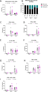
Fig 3. Ilomastat does not alter the inflammatory response to IAV.
Male and female wild-type C57BL/6 mice were instilled with PR8 or PBS in the presence or absence of 100mg/kg ILO or vehicle control administered intra-peritoneally daily from the time of infection. Samples were collected at day 1, 3, and 6. ILO treatment did not alter the (A) number or (B) composition of immune cells in BAL and did not significantly alter the BAL concentration of (C) Interferon (IFN)-α, (D) IFN-γ, (E) Interleukin (IL)-1β, (F) IL-6, (G) IL-10, (H) TNF-α, or (I) CXCL1/KC during IAV (n = 6–9 [4 male, 5 female], two-way ANOVA). There are no statistically significant sex-specific differences in these parameters.
Fig 4
Fig 4. Destruction of the glycocalyx in epithelial cells increases IAV viral load.
Confluent MLE-12 murine epithelial cells were treated with active or heat-inactivated heparinase I/III for 6h to shed the glycocalyx before infecting with PR8 at an MOI of 1 for 24 hours. (A) Destruction of the glycocalyx by heparinase I/III leads to increased viral load after PR8 infection (n = 12–17, overall p<0.0001 for infection, overall p = 0.025 for heparinase treatment, two-way ANOVA with significant post-hoc relationships between groups indicated). (B) MMP-7 and (C) TIMP-1 protein are not significantly altered by heparinase treatment (n = 11, two-way ANOVA), nor are (D-J) the concentration of several pro-inflammatory cytokines in the supernatant (n = 9–11, two-way ANOVA). For (G) IFNα, (H) IFNγ, (I) IL-1β, and (J) IL-10, samples are at or near the limit of detection.
Fig 5
Fig 5. MMP-7 and its inhibitor TIMP-1 have differential RNA localization with IAV genome.
On day 6 post infection, (A) MMP-7 and (B) TIMP-1 RNA expression in total lung lysates was increased by infection but not by ILO treatment (n = 5–12 [all male], MMP-7 overall p = 0.0086 for infection, TIMP-1 overall p = 0.0038 for infection, two-way ANOVAs). MMP-7 expression is limited to cells directly infected with IAV in (C) large, proximal airways at day 3 (n = 3 [all male], ≥9 infected fields per mouse) and in (D) smaller, distal airways at day 5 (n = 3–4 [all male], ≥9 infected fields per mouse). TIMP-1 expression is limited to cells adjacent but not directly infected with IAV at (E) day 3 (n = 4 [all male], ≥6 infected fields per mouse) and (F) day 7 (n = 4 [all male], ≥6 infected fields per mouse). IAV genome segment 8 and MMP-7 or TIMP-1 localization in whole lung tissue slices is visualized by in situ RNA hybridization, with 40x image fields representative of IAV and PBS mice shown. (G) There is no significant difference in MMP-7 protein in whole lung homogenates during IAV infection (n = 4–11 [7 male, 4 female], two-way ANOVA). (H) TIMP-1 total protein in whole lung homogenates increases on day 3 and day 7 after IAV (n = 6–11 [7 male, 4 female], overall p<0.0001 for infection, overall p<0.0001 for time, two-way ANOVA with significant post-hoc relationships within days indicated).
Fig 6
Fig 6. MMP-7 is not required for glycocalyx shedding, barrier permeability, or inflammation during IAV infection.
MMP-7KO or WT littermate control C57BL/6 were intranasally administered PR8 influenza or PBS and monitored for up to 7 days. MMP-7KO mice had no significant difference in (A) glycocalyx shedding (n = 4–11 [5 male, 6 female]; two-way ANOVA), (B) BAL protein content (n = 4–11 [5 male, 6 female], two-way ANOVA), or (C) weight loss compared to WT controls (n = 4–11 [5 male, 6 female], mixed model ANOVA). MMP-7KO mice did not have significantly altered (D) number or (E) composition of inflammatory cells present in the lung on day 6. (n = 4–11 [5 male, 6 female], two-way ANOVA). (F) MMP-7KO mice have similar viral load to WT littermates as seen by ΔΔCt analysis to same day WT control (n = 4–11 [5 male, 6 female], two-way ANOVA). There are no statistically significant sex-specific differences in these parameters.

References

    1. Center for Disease Control and Prevention. Past Seasons Estimated Influenza Disease Burden 2024 [https://www.cdc.gov/flu/about/burden/past-seasons.html.
    1. Taubenberger JK, Morens DM. The pathology of influenza virus infections. Annu Rev Pathol. 2008;3:499–522. doi: 10.1146/annurev.pathmechdis.3.121806.154316 - DOI - PMC - PubMed
    1. Timm S, Lettau M, Hegermann J, Rocha ML, Weidenfeld S, Fatykhova D, et al. The unremarkable alveolar epithelial glycocalyx: a thorium dioxide-based electron microscopic comparison after heparinase or pneumolysin treatment. Histochem Cell Biol. 2023;160(2):83–96. doi: 10.1007/s00418-023-02211-7 - DOI - PMC - PubMed
    1. Rizzo AN, Schmidt EP. The role of the alveolar epithelial glycocalyx in acute respiratory distress syndrome. Am J Physiol Cell Physiol. 2023;324(4):C799–C806. doi: 10.1152/ajpcell.00555.2022 - DOI - PMC - PubMed
    1. Ochs M, Hegermann J, Lopez-Rodriguez E, Timm S, Nouailles G, Matuszak J, et al. On Top of the Alveolar Epithelium: Surfactant and the Glycocalyx. Int J Mol Sci. 2020;21(9). - PMC - PubMed

MeSH terms