. 2025 May 1;64(5):3125-3133.
doi: 10.1093/rheumatology/keae517.

Mycophenolate mofetil directly modulates myeloid viability and pro-fibrotic activation of human macrophages

Affiliations

Mycophenolate mofetil directly modulates myeloid viability and pro-fibrotic activation of human macrophages

Emily A Morris et al. Rheumatology (Oxford). .

Abstract

Objectives: Mycophenolate mofetil (MMF) is an immunosuppressant used to treat rheumatological diseases, including systemic sclerosis (SSc). While MMF is an established inhibitor of lymphocyte proliferation, recent evidence suggests MMF also mediates effects on other cell types. The goal of this study was to determine the effect of MMF on monocytes and macrophages, which have been implicated in SSc pathogenesis.

Methods: Human monocyte-derived macrophages were cultured with the active MMF metabolite, mycophenolic acid (MPA), and assessed for changes in viability and immuno-phenotype. Guanosine supplementation studies were performed to determine whether MPA-mediated effects were dependent on de novo purine synthesis. The ability of MPA-treated macrophages to induce fibroblast activation was evaluated, and dermal myeloid expression signatures were analysed in MMF-treated SSc patients.

Results: MPA reduced viability and induced apoptosis in monocytes and macrophages at doses (average IC50 = 1.15 µg/ml) within the target serum concentration of MMF-treated SSc patients (1-3 µg/ml). These effects were reversed by guanosine supplementation. Low-dose MPA (0.5 µg/ml) attenuated IL-4 or SSc plasma-mediated macrophage activation, and inhibited the ability of SSc plasma-activated macrophages to induce SSc fibroblast activation. Gene expression studies demonstrated significant reductions in dermal myeloid signatures in MMF-responsive SSc patients.

Conclusion: For the first time, we have demonstrated that MMF inhibits the viability and pro-fibrotic activation of human monocytes and macrophages, which is dependent on de novo purine synthesis. Coupled with myeloid gene expression attenuation following MMF treatment in patients, these results suggest that the fibrotic inhibition observed with MMF may be attributable, at least in part, to direct effects on myeloid cells.

Keywords: fibrosis; inflammation; macrophages; mycophenolate mofetil; systemic sclerosis.


Figures

Figure 1.
Figure 1.
Mycophenolic acid (MPA) reduced viability and induced apoptosis in human monocytes and macrophages. (A) Peripheral blood-derived human monocytes were cultured in complete Roswell Park Memorial Institute (RPMI) 1640 media containing M-CSF (20 ng/ml) and incubated with indicated doses of MPA. Viability was assessed using the CellTiter Blue assay for mitochondrial function. (B–D) Monocyte-differentiated macrophages grown in 2 µg/ml MPA (approximate IC50; half maximal inhibitory concentration) were assessed for viability and induction of apoptosis via CellTiter Blue (C) and Caspase-Glo™ (D), respectively. Caspase activity luminescence values were normalized to the % of viable cells. Significance was determined by one-way ANOVA. n = 4 biological replicates; an average of n = 4 technical replicates is shown. Figures are representative of at least three independent experiments. Individual dose and time point comparison analysis can be found in Supplementary Fig. S1, available at Rheumatology online. ANOVA: analysis of variance
Figure 2.
Figure 2.
Low-dose mycophenolic acid (MPA) impaired the ability of IL-4 to activate human macrophages. (A) Low-dose MPA (0.5 µg/ml) or vehicle (dimethyl sulfoxide, DMSO) was cultured with human monocytes in complete Roswell Park Memorial Institute (RPMI) media and M-CSF (20 ng/ml). Cells were differentiated in the presence of MPA or vehicle for 72 h prior to activation with 20 ng/ml IL-4 and harvested for mRNA extraction or flow cytometry. (B) mRNA levels of C-C motif chemokine ligand 2 (CCL2), IL-10 and CD206 of IL-4–activated macrophages treated ± MPA; n = 3 biological replicates × n = 3 technical replicates. (C) MFI for surface expression of CD206 in MPA or control-treated cells measured using flow cytometry; representative of three biological replicates with n = 3 technical replicates each. The gating strategy can be found in Supplementary Fig. S2, available at Rheumatology online; only the populations of live singlet macrophages were used for MFI calculations. (D) ELISA quantification of secreted CCL2 in supernatants from IL-4–activated macrophages that were incubated with MPA (0.5 mg/ml) or vehicle. Data shown are representative of three biological replicates with n = 3 technical replicates. Significance was determined by Student’s t test; *P < 0.05, **P < 0.01, ***P < 0.001, ns: not significant
Figure 3.
Figure 3.
MPA effects on myeloid cells were IMPDH pathway-dependent. (A) Diagram of MPA inhibition of purine synthesis and salvage pathway (R5P, ribose-5-phosphate; PRPP, 5-phospho-α-D-ribosyl 1-pyrophosphate; IMP, inosine monophosphate; XMP, xanthine monophosphate; GMP, guanosine monophosphate; GDP, guanosine diphosphate; GTP, guanosine triphosphate; HPRT, hypoxanthine phosphoribosyltransferase; IMPDH, inosine monophosphate dehydrogenase; BH4, tetrahydrobiopterin; iNOS, inducible nitric oxide synthase; ROS, reactive oxygen species; MPA, mycophenolic acid; SAICAR, Phosphoribosylaminoimidazolesuccinocarboxamide; AICAR, 5-Aminoimidazole-4-carboxamide ribonucleotide; sAMP, adenylosuccinate; ADP, adenosine diphosphate; ATP, adenosine triphosphate; AMP, adenosine monophosphate; XO, xanthine oxidase). Guanosine (GUO) and guanine are produced downstream of IMPDH, and can be restored by salvage pathway activity (which is absent in lymphocytes, the primary target cells of MPA, but assumed to be present in myeloid cells). Supplementation with guanosine counters IMPDH-enzyme inhibition by restoring GTP and guanine availability. (B) MPA dose response curve for human monocyte-derived macrophages in the presence of 100μM guanosine. (C-D) Human monocyte-derived macrophages were cultured for 72 hours in 0, 2, or 4μg/mL MPA +/- 100μM guanosine and assessed for viability (C) and induction of apoptosis (D). Caspase-Glo™ values were normalized to the % of viable cells. (E) Monocytes were cultured in complete RPMI media with 20 ng/ml M-CSF and treated with low dose MPA (0.5μg/mL) and/or guanosine (GUO; 100μM)(DMSO, dimethyl sulfoxide was used for vehicle control). IL-4 was added after 72 hours to macrophages with or without MPA and guanosine. Viability and surface levels of cluster of differentiation (CD)206, human mannose receptor) were assessed via flow cytometry. Gating strategy can be found in Supplemental Figure S2, available at Rheumatology online; live singlet macrophages were used for MFI (mean fluorescence intensity) calculations. An average of 4 technical replicates for n = 4 biological replicates were analyzed in B–D; MFI values are representative of at least 3 biological replicates. Significance was assessed by student’s t-test or one-way ANOVA (analysis of variance); *P < 0.05, **P < 0.01, ***P < 0.001, ****P < 0.0001, n.s: non-significant
Figure 4.
Figure 4.
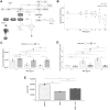
Low-dose MPA inhibited SSc plasma activation of human macrophages. Monocytes were differentiated in complete Roswell Park Memorial Institute (RPMI) media with low-dose mycophenolic acid (MPA) (0.5 µg/ml) or vehicle (DMSO) for 72 h. Media were then replaced with RPMI and SSc donor plasma, ± MPA for 48 h; average values for n = 3 technical replicates for n = 3 biological replicates are shown. (A) MFI (mean fluorescence intensity) for cluster of differentiation (CD)206, cluster of differentiation (CD)163 and human leukocyte antigen-DR (HLA-DR) were analysed using flow cytometry. The gating strategy is shown in Supplementary Fig. S4A, available at Rheumatology online. (B) mRNA expression of CD206, CD163, C-C motif chemokine ligand 2 (CCL2), IL-6 and IL-10 were assessed by quantitative real time polymerase chain reaction (qRT-PCR). Average values of n = 2 technical replicates for n = 3 biological replicates are presented. (C–D) Protein levels of CCL2 and IL-6 in macrophage supernatants were analysed by ELISA. (E) Experimental design for fibroblast activation experiment: SSc patient fibroblasts were seeded at 20 000 cells/well in 24-well plates in fibroblast growth media (FGM). After 48 h, media were replaced with a 1:1 dilution of fresh FGM and conditioned media (CM) from macrophages treated with vehicle (CM) or low-dose MPA (0.5 mg/ml, CM+MPA) prior to/during incubation with SSc plasma for 48 h. Fibroblasts were cultured with a 1:1 dilution of fresh FGM to CM from macrophages treated with or without MPA for 48 h, followed by total RNA isolation. Fibroblasts were cultured with CM or CM+MPA for 48 h, followed by total RNA isolation. (F) mRNA expression of alpha smooth muscle actin (αSMA), fibronectin 1 (FN1), collagen1A2 (COL1A2), and collagen3A1 (COL3A1) in SSc fibroblasts cultured with CM or CM+MPA were assessed by qRT-PCR; average values of n = 3 technical replicates for n = 3 biological replicates are shown. Significance was determined by paired Student’s t test; *P < 0.05, **P < 0.01, ***P < 0.001, ****P < 0.0001
Figure 5.
Figure 5.
Changes in the myeloid-specific gene signature during mycophenolate mofetil (MMF) treatment in dcSSc skin. (A) The expression levels of genes associated with the monocyte/myeloid cell signature in skin biopsies of five SSc patients, both at baseline (pre-MMF or MMF-naïve) and 24 months following MMF therapy (post-MMF) [6]. (B) Gene Set Variation Analysis (GSVA) enrichment score of monocyte/myeloid cell signature at baseline and 24 months after MMF therapy. (C) CCL2 and MRC1(CD206) expression levels at baseline (pre-MMF) and 24 months post-MMF treatment. (D) Changes in expression of the monocyte/myeloid cell signature of three dcSSc subjects whose MMF treatment was discontinued after 24 months. (E) GSVA enrichment score of monocyte/myeloid cell signature pre and post-MMF discontinuation. (F) CCL2 and MRC1(CD206) expression rates in patients before and after MMF discontinuation. Significance was determined by Student’s t test; *P < 0.05, **P < 0.01, ***P < 0.001

References

    1. Allison AC, Eugui EM. Mycophenolate mofetil and its mechanisms of action. Immunopharmacology 2000;47:85–118. - PubMed
    1. Bernstein EJ, Huggins JT, Hummers LK, Owens GM. EBSCOhost | 157543047 | systemic sclerosis with associated interstitial lung disease: management considerations and future directions. Am J Manag Care 2021;27:S138–46. - PubMed
    1. Mehling A, Grabbe S, Voskort M et al. Mycophenolate mofetil impairs the maturation and function of murine dendritic cells. J Immunol 2000;165:2374–81. - PubMed
    1. Allison AC, Eugui EM. Mechanisms of action of mycophenolate mofetil in preventing acute and chronic allograft rejection. Transplantation 2005;80:S181–90. - PubMed
    1. Volkmann ER, Andréasson K, Smith V. Systemic sclerosis. Lancet 2022;401:304–18. - PMC - PubMed

MeSH terms