. 2024 Oct;121(40):e2402368121.
doi: 10.1073/pnas.2402368121. Epub 2024 Sep 23.

Nr2f1 enhancers have distinct functions in controlling Nr2f1 expression during cortical development

Affiliations

Nr2f1 enhancers have distinct functions in controlling Nr2f1 expression during cortical development

Zhidong Liu et al. Proc Natl Acad Sci U S A. 2024 Oct.

Abstract

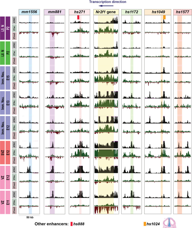
There is evidence that transcription factor (TF) encoding genes, which temporally control development in multiple cell types, can have tens of enhancers that regulate their expression. The NR2F1 TF developmentally promotes caudal and ventral cortical regional fates. Here, we epigenomically compared the activity of Nr2f1's enhancers during mouse cortical development with their activity in a transgenic assay. We identified at least six that are likely to be important in prenatal cortical development, with three harboring de novo mutants identified in ASD individuals. We chose to study the function of two of the most robust enhancers by deleting them singly or together. We found that they have distinct and overlapping functions in driving Nr2f1's regional and laminar expression in the developing cortex. Thus, these two enhancers, probably in combination with the others that we defined epigenetically, precisely tune Nr2f1's regional, cell type, and temporal expression during corticogenesis.

Keywords: Nr2f1; cortical development; enhancer; epigenomics.


Conflict of interest statement

Competing interests statement:J.L.R. is cofounder, stockholder, and currently on the scientific board of Neurona, a company studying the potential therapeutic use of interneuron transplantation. J.L.R has stock in Neurona.

Figures

Fig. 1.
Fig. 1.
Nr2f1 telencephalic enhancers. Selected Nr2f1 enhancers show telencephalic activity closely resembling Nr2f1 expression at E11.5. (A) Schema showing a 4 Mb genomic region surrounding the Nr2f1 locus (mm10, chr13:76762978-80762978) (red bar: Nr2f1 gene body. (B) Bar graph showing 35 Vista enhancers around the Nr2f1 locus; 8 are active in the telencephalon (red bars). 15 are inactive (black bars), 7 are active but not in the forebrain (green bars), and 5 are active in the forebrain but not in the telencephalon (blue bars) (Dataset S1). (C) 6 enhancers driving LacZ activity (blue) in the mouse pallium; whole mounts at E11.5 (a-g). These embryos were coronally sectioned; representative sections show LacZ activity in telencephalic regions. Because of its low background, we highlighted the circumference of mm881’s sections in black. In situ hybridization of Nr2f1 RNA (D1-D3). Black arrows indicate regions with either LacZ activity or Nr2f1 RNA. DP: dorsal pallium, LP: lateral pallium, VP: ventral pallium, MP; medial pallium, LGE: lateral ganglionic eminence, MGE: medial ganglionic eminence, CGE: caudal ganglionic eminence, Th: thalamus, POA: preoptic area. Magnification bar: 0.5 mm.
Fig. 2.
Fig. 2.
Nr2f1 locus organization and epigenetic analyses at E12.5. (A) The location of six pallial Nr2f1 VISTA enhancers (red) and their genomic distance to Nr2f1 gene are shown in the WashU genome browser. Other Nr2f1 VISTA enhancers are indicated in blue. The location of other genes is shown to the right of RefSq gene. (B) EMX2, LHX2, NR2F1, PAX6, and PBX1 TF ChIP-seq data in E12.5 cortex are displayed (black rectangles surround called peaks) (2). (C) Epigenetic (H3K27ac, H3K27me3, and ATAC) modifications to enhancers in the E12.5 cortex are shown for wildtype and Pax6−/−, Emx2−/−, and Nr2f1−/−(2). Red rectangles indicate gain of peak height in the mutants and green rectangles indicate loss of peak heights in the mutants. (D) Plac-seq data shows enhancer interactions with Nr2f1 promoter (2). (E) Locations of CRISPR deletions of hs271 and hs1172 enhancers are shown as red bars. Abbreviations: 27ac: H3K27ac, 27me3: H3K27me3, Mut: mutant. WT: wild type, pRE: putative regulatory element.
Fig. 3.
Fig. 3.
Developmental course of epigenetic states for six Nr2f1 pallial enhancers and Nr2f1 gene body in cortical VZ, SVZ, and immature neurons and deep layer neurons. ATAC-seq (ATAC in gray), H3K27ac (27ac in green), and H3K27me3 (27me3 in red) epigenetic marks are shown in cortical cell subtypes at different developmental ages. Each row represents a distinct cortical cell type at a specific age including VZ, SVZ, prenatal Immature Neurons, P2 Layer 6 Neurons (L6N), and P2 Layer 5 Neurons (L5N) (specified in text boxes on the left of the figure). Colored columns represent the genomic regions of each pallial enhancer as well as the Nr2f1 gene body (specified in text boxes at the top of the figure); blue for mm1556, purple for mm881, pink for hs271, yellow for Nr2f1 gene, green for hs1172, orange for hs1049, red for hs1577. Two additional VISTA enhancers with genomic coordinates close to the pallial enhancers are shown in the L5N row: hs888 in red near hs271 whose region of activity is unknown and hs1024 in orange near hs1049 with activity in intertelencephalic mesenchyme at E11.5 (see sectioned brain at the bottom of hs1049 column). The genomic region shown for each enhancer and the Nr2f1 gene body is 50 kb.
Fig. 4.
Fig. 4.
Changes in Nr2f1 pallial and subpallial expression in hs1172, hs271, and hs1172/ hs271 mutants at E12.5. Nr2f1 in situ RNA expression in telencephalic coronal hemisections (rostral to caudal) in WT (AA4), hs1172−/− (BB4), hs271−/−(CC4), and hs1172−/−/ hs271−/−(DD4) at E12.5. NR2F1 immunochemistry in WT and mutants (A5–D5). Black arrows indicate pallial locations where Nr2f1 is expressed in WT. (E and F) Quantitative analysis of Nr2f1 expression levels in the lateral and dorsal rostral pallium. Purple rectangles indicated the areas where Nr2f1 in situ signal density was measured. Measurements were made in four brain sections using ImageJ. *P < 0.05, **P < 0.01, ***P < 0.001, ****P < 0.0001 (mean ± SD). Between the two green arrowheads are pallial regions with reduced Nr2f1 expression. Red arrowheads indicate reduced Nr2f1 expression in the pallial VZ. Orange arrowheads indicate reduced Nr2f1 expression in LGE, MGE, and CGE. Blue arrowheads indicate preserved Nr2f1 expression in a superficial layer of the pallium. Blue arrowheads also indicate preserved NR2F1 expression in a superficial layer of the pallium (A5–D5). Abbreviations: CP, cortical plate; OE, olfactory epithelium; Hyp: hypothalamus; for the rest of the abbreviations, see legend to Fig. 1. Magnification bar: 0.5 mm.
Fig. 5.
Fig. 5.
Changes in Nr2f1 pallial and subpallial expression in hs1172, hs271, and hs1172/ hs271 mutants at E14.5. Nr2f1 in situ RNA expression in telencephalic coronal hemisections (rostral to caudal) in WT (AA4), hs1172−/− (BB4), hs271−/−(CC4), and hs1172−/−/ hs271−/−(DD4). NR2F1 immunochemistry in WT and mutants (A5–D5) at E14.5. Black arrows indicate pallial locations where Nr2f1 is expressed in WT. Between the two green arrowheads are pallial regions with reduced Nr2f1 expression. Red arrowheads indicate reduced Nr2f1 expression in the pallial VZ. Orange arrowheads indicate reduced Nr2f1 expression in LGE, MGE, and CGE. Blue arrowheads indicate preserved Nr2f1 expression in the pallial SVZ of the hs271−/−and hs1172−/−/ hs271−/−despite the reduction in the VZ. Blue arrowheads also indicate preserved NR2F1 expression in the pallial SVZ of the hs271−/−and hs1172−/−/ hs271−/−(A5–D5). Abbreviations: PTh: prethalamus; for the rest of the abbreviations, see legends to Figs. 1 and 4. Magnification bar: 0.5 mm.
Fig. 6.
Fig. 6.
Deletion of Nr2f1 enhancers results in a caudal expansion of rostral cortical properties marked by NT3. NT3 in situ RNA expression in telencephalic coronal hemisections (rostral to caudal) in WT (AA5), hs1172−/− (BB5), hs271−/−(CC5), and hs1172−/−/ hs271−/−(DD5) at P5. NT3 strongly marks the PFC (lateral and dorsal parts), Cg and RS, is weak in the MCx, and not detectable in the SSCx (delimited by two green arrowheads). hs1172−/− shows no phenotype, whereas the hs271−/−and hs1172−/−/ hs271−/− lack the NT3-negative SSCx area in rostral sections. Abbreviation: Mos/mdPFC: secondary motor area/dorsomedial prefrontal cortex, vmPFC: ventromedial prefrontal cortex, AI: agranular insular, ORBI: orbital areas, ICx: insular cortex, SSCx: somatosensory cortex, MCx: motor cortex; Cg: cingulate cortex, RS: retrosplenial cortex. Magnification bar: 0.5 mm.
Fig. 7.
Fig. 7.
Lmo4 expression patterning showed Nr2f1 enhancers mutants had rostral/caudal cortical reginal patterning defects. Lmo4 in situ RNA expression in telencephalic coronal hemisections (rostral to caudal) in WT (AA6), hs1172−/− (BB6), hs271−/−(CC6), and hs1172−/−/ hs271−/−(DD6) at P5. Lmo4 marks the PFC (lateral, dorsal, medial and orbital parts), MCx, Cg, RS, VCx, and ACx but is weak in the SSCx (delimited by two green arrowheads). hs1172−/− shows no phenotype in rostral cortex, whereas caudally there is an expansion of the Lmo4-negative SSCx area. hs271−/−and hs1172−/−/ hs271−/− lack the Lmo4-negative SSCx area in rostral sections, caudally there is a robust expansion of the Lmo4-negative SSCx area. Abbreviations: ACx: auditory cortex, for the rest of the abbreviations, see legend to Fig. 6. Magnification bar: 0.5 mm.
Fig. 8.
Fig. 8.
De novo mutations in VISTA elements near human NR2F1. VISTA regulatory elements (purple) and genes (blue) in the genomic region around NR2F1 are shown at the bottom. PhyloP conservation scores for the region based on 100-way vertebrate analysis are shown under the genes. Three inserts show the location of seven de novo mutations observed in 8,626 ASD cases (red) and 4,461 sibling controls (blue). Labels show the sample size, genomic position of the first nucleotide, and the nucleotide change (VCF format).

Similar articles

References

    1. Chen B., Kwan K. Y., Rubenstein J. L. R., Rakic P., Patterning and Cell Type Specification in the Developing CNS and PNS: Comprehensive Developmental Neuroscience (Academic Press, Amsterdam, The Netherlands, ed. 2, 2020), pp. xxiv, 1098pp.
    1. Ypsilanti A. R., et al. , Transcriptional network orchestrating regional patterning of cortical progenitors. Proc. Natl. Acad. Sci. U.S.A. 118, e2024795118 (2021). - PMC - PubMed
    1. Visel A., Minovitsky S., Dubchak I., Pennacchio L. A., VISTA Enhancer Browser–a database of tissue-specific human enhancers. Nucleic Acids Res. 35, D88–D92 (2007). - PMC - PubMed
    1. Visel A., et al. , A high-resolution enhancer atlas of the developing telencephalon. Cell 152, 895–908 (2013). - PMC - PubMed
    1. Bejerano G., et al. , Ultraconserved elements in the human genome. Science 304, 1321–1325 (2004). - PubMed

Substances

LinkOut - more resources