. 2024 Oct;121(40):e2402983121.
doi: 10.1073/pnas.2402983121. Epub 2024 Sep 23.

A sensitive assay for measuring whole-blood responses to type I IFNs

Affiliations

A sensitive assay for measuring whole-blood responses to type I IFNs

Adrian Gervais et al. Proc Natl Acad Sci U S A. 2024 Oct.

Abstract

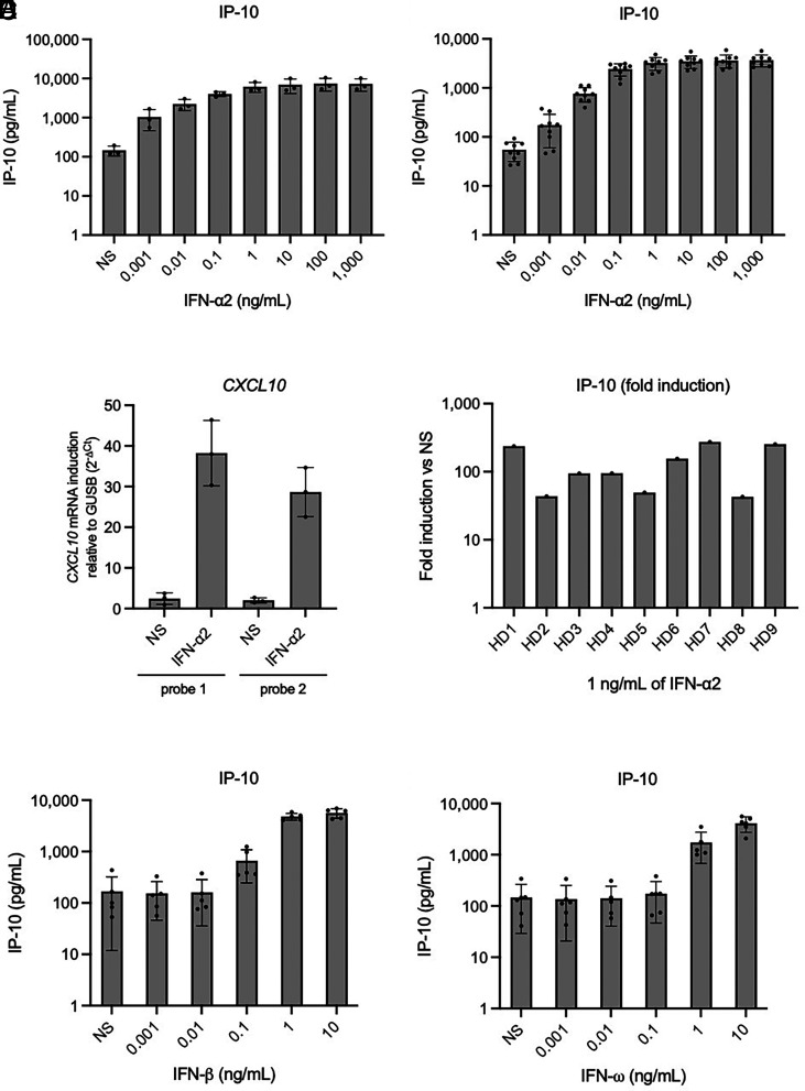
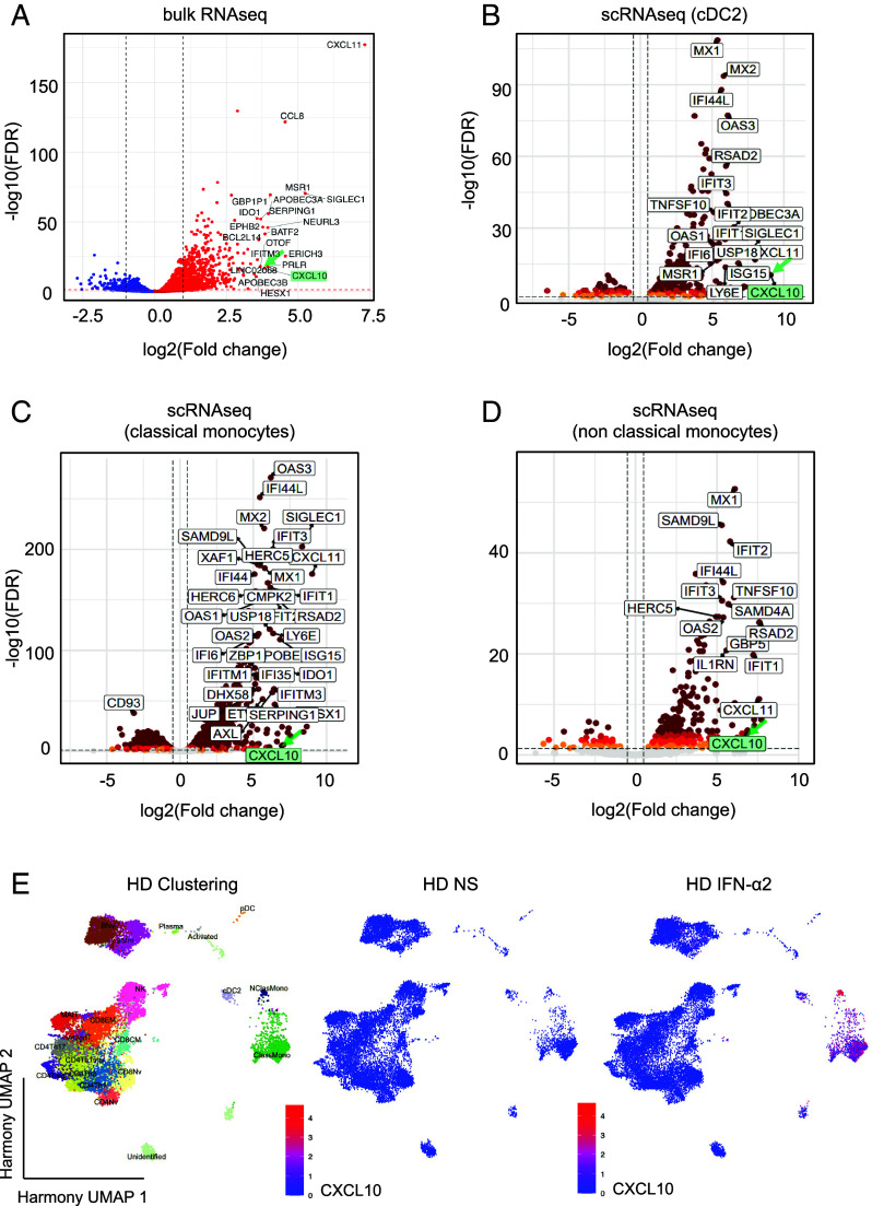
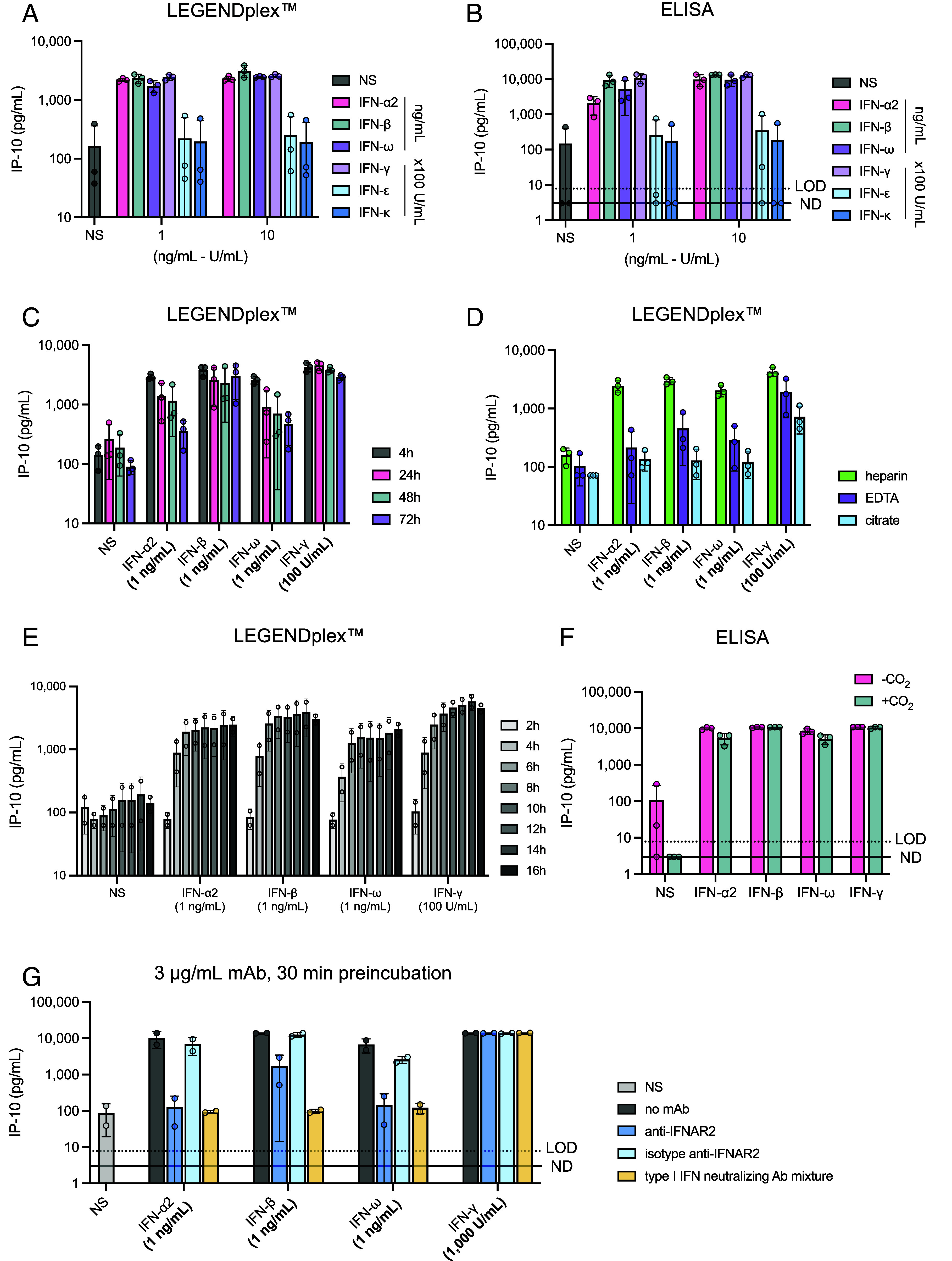
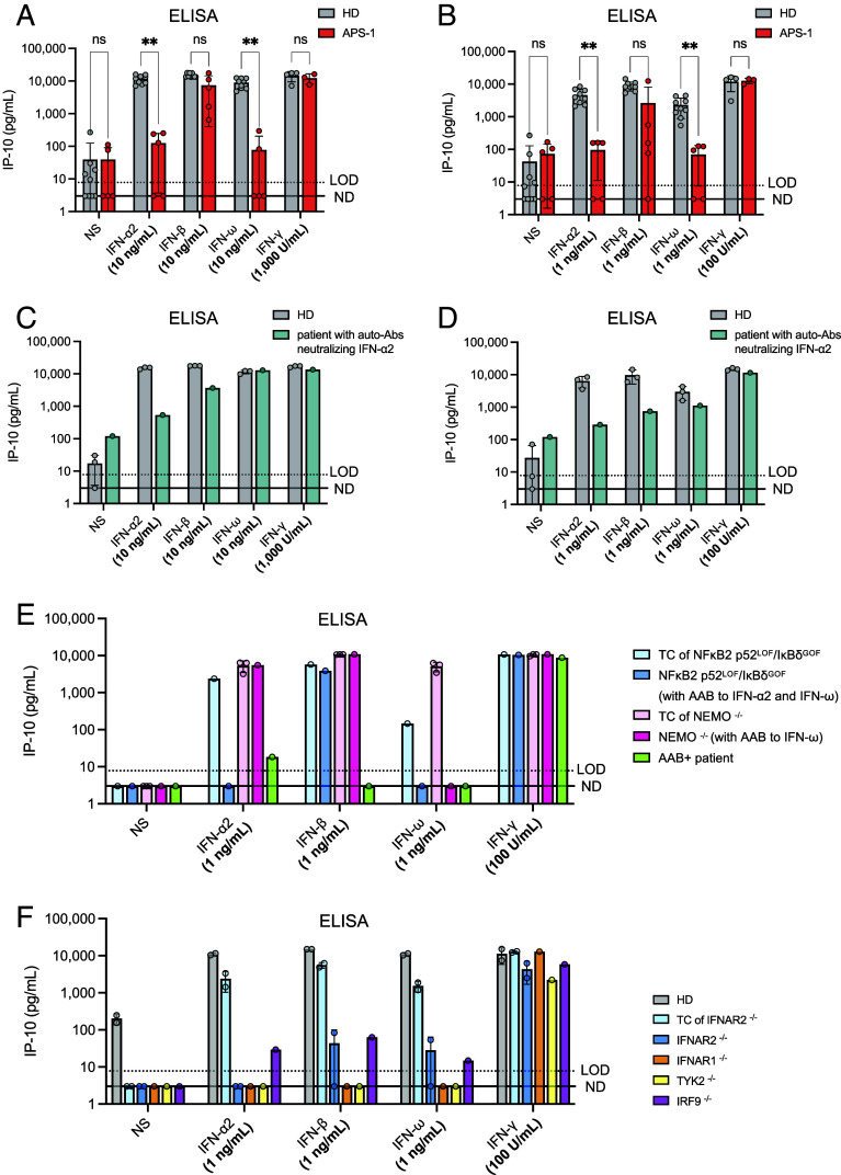
Human inborn errors of the type I IFN response pathway and auto-Abs neutralizing IFN-α, -β, and/or -ω can underlie severe viral illnesses. We report a simple assay for the detection of both types of condition. We stimulate whole blood from healthy individuals and patients with either inborn errors of type I IFN immunity or auto-Abs against type I IFNs with glycosylated human IFN-α2, -β, or -ω. As controls, we add a monoclonal antibody (mAb) blocking the type I IFN receptors and stimulated blood with IFN-γ (type II IFN). Of the molecules we test, IP-10 (encoded by the interferon-stimulated gene (ISG) CXCL10) is the molecule most strongly induced by type I and type II IFNs in the whole blood of healthy donors in an ELISA-like assay. In patients with inherited IFNAR1, IFNAR2, TYK2, or IRF9 deficiency, IP-10 is induced only by IFN-γ, whereas, in those with auto-Abs neutralizing specific type I IFNs, IP-10 is also induced by the type I IFNs not neutralized by the auto-Abs. The measurement of type I and type II IFN-dependent IP-10 induction therefore constitutes a simple procedure for detecting rare inborn errors of the type I IFN response pathway and more common auto-Abs neutralizing type I IFNs.

Keywords: ELISA; IP-10; auto-antibodies; diagnostic test; type I interferons.


Conflict of interest statement

Competing interests statement:J.L.-C. is an inventor on patent application PCT/US2021/042741, filed July 22, 2021, submitted by The Rockefeller University and covering the diagnosis of susceptibility to, and the treatment of, viral disease, and viral vaccines, including COVID-19 and vaccine-associated diseases. Reviewer S.R.O. is a co-author of two recent papers with J.L.C. Reviewer P.H. has a collaboration with the Casanova group in the exchange of reagents, but it does not cover the topic of this collaboration.

Figures

Fig. 1.
Fig. 1.
IP-10 (CXCL10) induction after the stimulation of whole blood from healthy donors with type I IFNs. (A and B) IP-10 induction, assessed with plasma supernatants after the stimulation of whole blood with various concentrations of glycosylated IFN-α2 for 16 h, as measured by LEGENDplex™. Blood samples were collected 8 to 24 h before stimulation. Three (A) or nine (B) healthy donors were tested once for each set of conditions. (C) CXLC10 mRNA induction after the stimulation of PBMCs from three healthy donors with 1 ng/mL glycosylated IFN-α2 for 6 h, as measured by RT-qPCR. (D) Fold-induction of IP-10 after the stimulation of whole blood with 1 ng/mL glycosylated IFN-α2 for 16 h, as measured by LEGENDplex™. Nine healthy donors were tested once each. Blood samples were collected 8 to 24 h before stimulation. (E and F) IP-10 induction, assessed with plasma supernatants after the stimulation of whole blood with various concentrations of glycosylated IFN-β (E) or glycosylated IFN-ω (F) for 16 h, as measured by LEGENDplex™. Blood samples were collected 8 to 24 h before stimulation. Five healthy donors were tested once for each set of conditions.
Fig. 2.
Fig. 2.
Whole-transcriptome analysis after PBMC stimulation with IFN-α2. (A) Volcano plot analysis of bulk RNAseq performed on total mRNA extracted from the PBMCs of three healthy donors after stimulation with 1 ng/mL glycosylated IFN-α2 for 6 h. The labeled genes are the top 20 genes displaying the highest levels of induction relative to nonstimulated conditions. (BD) Volcano plot analysis showing the transcripts induced in cDC2 cells (B), classical monocytes (C), and nonclassical monocytes (D), as determined by scRNAseq (Parse Bioscience). PBMCs of three healthy donors were stimulated with 1 ng/mL glycosylated IFN-α2 for 6 h and whole mRNA was extracted. (E) Single-cell transcriptom analysis. PBMCs from three HD were analyzed after IFN-α2 stimulation vs unstimulated. Clustering analysis. After batch correction with Harmony, celltypes were identified manually. Feature plots show the induction of CXCL10 (encoding IP-10).
Fig. 3.
Fig. 3.
Evaluation of technical parameters and assessment of the activity of other cytokines (human IFN-ε, IFN-κ, and IFN-γ) on IP-10 induction. (A) IP-10 induction after stimulation with 100 U/mL or 1,000 U/mL recombinant human IFN-ε, IFN-κ, or IFN-γ for 16 h. Three healthy donors were tested for each set of conditions. IP-10 levels were then assessed in plasma supernatants by LEGENDplex™. (B) IP-10 induction after stimulation with 100 U/mL or 1,000 U/mL recombinant human IFN-ε, IFN-κ, or IFN-γ for 16 h. Three healthy donors were tested for each set of conditions. IP-10 levels were then assessed in plasma supernatants by ELISA. (C) Effect of time between collection and stimulation on IP-10 induction. IP-10 levels were assessed in plasma supernatants by LEGENDplex™ after the stimulation of whole blood with 1 ng/mL IFN-α2, -β, -ω, or -γ for 16 h. Three healthy donors were tested for each set of conditions. (D) Effect of the matrix on which blood is collected. Lithium heparin, EDTA, and citrate tubes were used to collect blood from three healthy donors. Whole-blood samples were stimulated with 1 ng/mL IFN-α2, -β, -ω, or -γ for 16 h. IP-10 levels were then assessed in plasma supernatants by LEGENDplex™. (E) Kinetics of IP-10 induction (effect of stimulation time). Whole blood from two healthy donors was stimulated with the indicated concentrations of IFNs for 2, 4, 6, 8, 10, 12, 14, and 16 h. IP-10 levels were then assessed in plasma supernatants by LEGENDplex™. (F) Effect of the presence or absence of 5% CO2 on IP-10 production after the stimulation of whole blood from three healthy donors with 1 ng/mL IFN-α2, -β, -ω, or -γ for 16 h. −CO2: stimulation at 37 °C without CO2 supplementation, +CO2: stimulation at 37 °C with 5% CO2 supplementation. (G) IP-10 induction after the stimulation of whole blood from two HDs with type I IFNs and with preincubation with 3 µg/mL anti-IFNAR2 neutralizing mAb, isotype antibody, or a cocktail of antibodies neutralizing all type I IFNs (dilution 1:50). The mAbs were incubated with whole blood for 30 min before stimulation with IFNs.
Fig. 4.
Fig. 4.
IP-10 induction after the stimulation of whole blood from patients with impaired type I IFN-dependent immunity. (A) IP-10 induction after the stimulation of whole blood from five APS-1 patients and nine healthy donors with 10 ng/mL glycosylated IFN-α2, IFN-β, or IFN-ω for 16 h. IP-10 levels were measured in plasma supernatants by ELISA. Whole blood from three APS-1 patients and five healthy donors was also stimulated with 1,000 U/mL IFN-γ as a control. IP-10 levels were compared between the HD and APS-1 groups by implementing nonparametric Mann–Whitney tests in GraphPad Prism. Ns: not significant; **P-value < 0.001. (B) IP-10 induction after the stimulation of whole blood from five APS-1 patients and nine healthy donors with 1 ng/mL glycosylated IFN-α2, IFN-β, or IFN-ω for 16 h. IP-10 levels were measured in plasma supernatants by ELISA. Whole blood from three APS-1 patients and five healthy donors was also stimulated with 100 U/mL IFN-γ as a control. Multiple Mann–Whitney tests were performed to compare the HD and APS-1 groups for each set of stimulation conditions. IP-10 levels were compared between the HD and APS-1 groups in nonparametric Mann–Whitney tests implemented in GraphPad Prism. Ns: not significant; **P-value < 0.001. (C) IP-10 induction in plasma supernatants after the stimulation of whole blood from a patient neutralizing low concentrations of IFN-α2 (1 ng/mL to 100 pg/mL) in a luciferase assay. Whole-blood samples from the patient and three healthy donors were stimulated with glycosylated type I (10 ng/mL) or type II (1,000 U/mL) IFNs for 16 h. IP-10 levels were then assessed in plasma supernatants by ELISA. (D) IP-10 induction in plasma supernatants after the stimulation of whole blood from a patient neutralizing low concentrations of IFN-α2 (1 ng/mL to 100 pg/mL) in a luciferase assay. Whole-blood samples from the patient and three healthy donors were stimulated with glycosylated type I (1 ng/mL) or type II (100 U/mL) IFNs for 16 h. IP-10 levels were then assessed in plasma supernatants by ELISA. (E) IP-10 induction in plasma supernatants after the stimulation of whole blood from a patient with a NFKB2 p52LOF/IκBδGOF mutation, a female patient with NEMO deficiency (incontinentia pigmenti), and a patient with auto-Abs neutralizing type I IFNs with no genetic diagnosis. TC: Travel control. Whole-blood samples were stimulated with glycosylated type I (1 ng/mL) or type II (100 U/mL) IFNs for 16 h. IP-10 levels were then assessed in plasma supernatants by ELISA. (F) IP-10 induction in plasma samples from patients with autosomal recessive IFNAR1, IFNAR2, TYK2, or IRF9 deficiency. Whole-blood samples from the patients and three healthy donors were stimulated with glycosylated type I (1 ng/mL) or type II (100 U/mL) IFNs for 16 h. IP-10 levels were then assessed in plasma supernatants by ELISA. A TC blood sample from a healthy individual was available for one of the IFNAR2−/− patients. These samples were stimulated >48 h after blood sampling. The blood of the TYK2−/− patient was stimulated 24 h after blood sampling. The blood of the IRF9−/− patient was stimulated 6 h after blood sampling.

Similar articles

Cited by

  • Auto-Abs neutralizing type I IFNs in patients with severe Powassan, Usutu, or Ross River virus disease.
    Gervais A, Bastard P, Bizien L, Delifer C, Tiberghien P, Rodrigo C, Trespidi F, Angelini M, Rossini G, Lazzarotto T, Conti F, Cassaniti I, Baldanti F, Rovida F, Ferrari A, Mileto D, Mancon A, Abel L, Puel A, Cobat A, Rice CM, Cadar D, Schmidt-Chanasit J, Scheid JF, Lemieux JE, Rosenberg ES, Agudelo M, Tangye SG, Borghesi A, Durand GA, Duburcq-Gury E, Valencia BM, Lloyd AR, Nagy A, MacDonald MM, Simonin Y, Zhang SY, Casanova JL. Gervais A, et al. J Exp Med. 2024 Dec 2;221(12):e20240942. doi: 10.1084/jem.20240942. Epub 2024 Nov 1. J Exp Med. 2024. PMID: 39485284 Free PMC article.
  • Autoantibodies neutralizing type I IFNs underlie severe tick-borne encephalitis in ∼10% of patients.
    Gervais A, Marchal A, Fortova A, Berankova M, Krbkova L, Pychova M, Salat J, Zhao S, Kerrouche N, Le Voyer T, Stiasny K, Raffl S, Schieber Pachart A, Fafi-Kremer S, Gravier S, Robbiani DF, Abel L, MacDonald MR, Rice CM, Weissmann G, Kamal Eldin T, Robatscher E, Erne EM, Pagani E, Borghesi A, Puel A, Bastard P, Velay A, Martinot M, Hansmann Y, Aberle JH, Ruzek D, Cobat A, Zhang SY, Casanova JL. Gervais A, et al. J Exp Med. 2024 Oct 7;221(10):e20240637. doi: 10.1084/jem.20240637. Epub 2024 Sep 24. J Exp Med. 2024. PMID: 39316018 Free PMC article.
  • HNMT Promotes the Occurrence and Progression of Nasopharyngeal Carcinoma by Inhibiting the IFN/TXNIP/p53 Axis.
    Cheng S, Wu XF, Sun WD, Zhai H, Liu X, Jiang CW, Ruan B. Cheng S, et al. Curr Med Sci. 2025 Jun;45(3):661-670. doi: 10.1007/s11596-025-00072-w. Epub 2025 Jun 11. Curr Med Sci. 2025. PMID: 40498422

References

    1. Pozzetto B., Mogensen K. E., Tovey M. G., Gresser I., Characteristics of autoantibodies to human interferon in a patient with varicella-zoster disease. J. Infect Dis. 150, 707–713 (1984), 10.1093/infdis/150.5.707. - DOI - PubMed
    1. Mogensen K. E., Daubas P. H., Gresser I., Sereni D., Varet B., Patient with circulating antibodies to α-interferon. Lancet 318, 1227–1228 (1981), 10.1016/S0140-6736(81)91460-4. - DOI - PubMed
    1. Casanova J.-L., Ion gresser. J. Interferon Cytokine Res. 39, 317–320 (2019), 10.1089/jir.2018.29015.mem. - DOI
    1. Levin M., Anti-interferon auto-antibodies in autoimmune polyendocrinopathy syndrome type 1. PLoS Med. 3, e292 (2006), 10.1371/journal.pmed.0030292. - DOI - PMC - PubMed
    1. Meager A., et al. , Anti-interferon autoantibodies in autoimmune polyendocrinopathy syndrome type 1. PLoS Med. 3, e289 (2006), 10.1371/journal.pmed.0030289. - DOI - PMC - PubMed

MeSH terms

Grants and funding

LinkOut - more resources