Artificial Intelligence for Contrast-Enhanced Ultrasound of the Liver: A Systematic Review
- PMID: 39312896
- PMCID: PMC12129427
- DOI: 10.1159/000541540
Artificial Intelligence for Contrast-Enhanced Ultrasound of the Liver: A Systematic Review
Abstract
Introduction: The research field of artificial intelligence (AI) in medicine and especially in gastroenterology is rapidly progressing with the first AI tools entering routine clinical practice, for example, in colorectal cancer screening. Contrast-enhanced ultrasound (CEUS) is a highly reliable, low-risk, and low-cost diagnostic modality for the examination of the liver. However, doctors need many years of training and experience to master this technique and, despite all efforts to standardize CEUS, it is often believed to contain significant interrater variability. As has been shown for endoscopy, AI holds promise to support examiners at all training levels in their decision-making and efficiency.
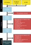
Methods: In this systematic review, we analyzed and compared original research studies applying AI methods to CEUS examinations of the liver published between January 2010 and February 2024. We performed a structured literature search on PubMed, Web of Science, and IEEE. Two independent reviewers screened the articles and subsequently extracted relevant methodological features, e.g., cohort size, validation process, machine learning algorithm used, and indicative performance measures from the included articles.
Results: We included 41 studies with most applying AI methods for classification tasks related to focal liver lesions. These included distinguishing benign versus malignant or classifying the entity itself, while a few studies tried to classify tumor grading, microvascular invasion status, or response to transcatheter arterial chemoembolization directly from CEUS. Some articles tried to segment or detect focal liver lesions, while others aimed to predict survival and recurrence after ablation. The majority (25/41) of studies used hand-picked and/or annotated images as data input to their models. We observed mostly good to high reported model performances with accuracies ranging between 58.6% and 98.9%, while noticing a general lack of external validation.
Conclusion: Even though multiple proof-of-concept studies for the application of AI methods to CEUS examinations of the liver exist and report high performance, more prospective, externally validated, and multicenter research is needed to bring such algorithms from desk to bedside.
Keywords: Artificial intelligence; Contrast-enhanced ultrasound; Hepatology; Liver; Sonography.
© 2024 The Author(s). Published by S. Karger AG, Basel.
Conflict of interest statement
J.N.K. declares consulting services for Owkin, France; DoMore Diagnostics, Norway; Panakeia, UK; Scailyte, Switzerland; Mindpeak, Germany; and MultiplexDx, Slovakia. Furthermore, he holds shares in StratifAI GmbH, Germany, has received a research grant by GSK, and has received honoraria by AstraZeneca, Bayer, Eisai, Janssen, MSD, BMS, Roche, Pfizer, and Fresenius. AB declares consulting services to Bracco and speaker’s fees from GE Healthcare and Hologic. M.K. declares honorary talks and travel support from Bracco Imaging and Canon Medical, and furthermore, he received a research grant by Bracco Imaging. No other conflicts of interest are declared by any of the authors.
Figures


References
-
- Center for Devices, Radiological Health . Artificial intelligence and machine learning (AI/ML)-Enabled medical devices [Internet]; US Food and Drug Administration. 2023. [cited 2024 Mar 14]. Available from: https://www.fda.gov/medical-devices/software-medical-device-samd/artific...
-
- Data IBM . AI vs. Machine learning vs. Deep learning vs. Neural networks: what’s the difference? [Internet]; IBM Blog. 2023. [cited 2024 Feb 27]. Available from: https://www.ibm.com/blog/ai-vs-machine-learning-vs-deep-learning-vs-neur...
-
- Mohri M, Rostamizadeh A, Talwalkar A. Foundations of machine learning. 2nd ed.MIT Press; 2018.
-
- StatQuest with Josh Starmer [Internet] . YouTube. [cited 2024 Apr 9]. Available from: https://www.youtube.com/channel/UCtYLUTtgS3k1Fg4y5tAhLbw
Publication types
MeSH terms
Substances
LinkOut - more resources
Full Text Sources
Medical
Research Materials

