. 2025 Jan;31(1):245-257.
doi: 10.1038/s41591-024-03309-8. Epub 2024 Sep 23.

Posthospitalization COVID-19 cognitive deficits at 1 year are global and associated with elevated brain injury markers and gray matter volume reduction

Greta K Wood #  1   2 Brendan F Sargent #  1   3   4 Zain-Ul-Abideen Ahmad  5 Kukatharmini Tharmaratnam  6 Cordelia Dunai  1   7 Franklyn N Egbe  1 Naomi H Martin  5 Bethany Facer  8 Sophie L Pendered  1 Henry C Rogers  5 Christopher Hübel  5   9   10 Daniel J van Wamelen  11   12   13 Richard A I Bethlehem  14 Valentina Giunchiglia  15 Peter J Hellyer  11 William Trender  15 Gursharan Kalsi  5 Edward Needham  16 Ava Easton  1   17 Thomas A Jackson  18 Colm Cunningham  19 Rachel Upthegrove  20 Thomas A Pollak  21   22 Matthew Hotopf  23 Tom Solomon  1   7   24   25 Sarah L Pett  26   27   28 Pamela J Shaw  29   30 Nicholas Wood  31   32 Neil A Harrison  33 Karla L Miller  4 Peter Jezzard  4 Guy Williams  34 Eugene P Duff  35 Steven Williams  11 Fernando Zelaya  11   36 Stephen M Smith  4 Simon Keller  8 Matthew Broome  20   37 Nathalie Kingston  38   39 Masud Husain  40   41 Angela Vincent  40 John Bradley  38 Patrick Chinnery  16   42 David K Menon  43 John P Aggleton  44 Timothy R Nicholson  21   22   45 John-Paul Taylor  46   47 Anthony S David  48 Alan Carson  49 Ed Bullmore  34   50 Gerome Breen  5   36 Adam Hampshire  11   15 COVID-CNS ConsortiumBenedict D Michael  51   52   53 Stella-Maria Paddick  46   54   55 E Charles Leek  56   57
Collaborators, Affiliations

Posthospitalization COVID-19 cognitive deficits at 1 year are global and associated with elevated brain injury markers and gray matter volume reduction

Greta K Wood et al. Nat Med. 2025 Jan.

Abstract

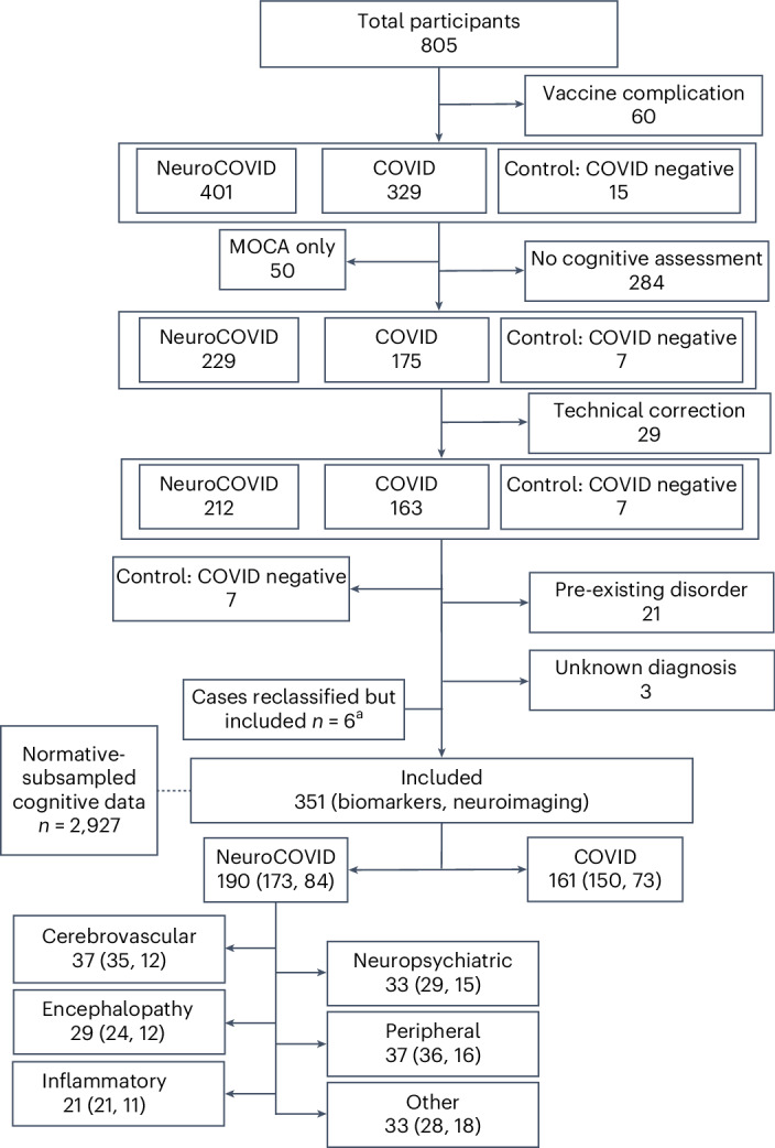
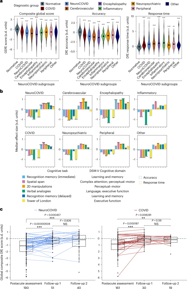
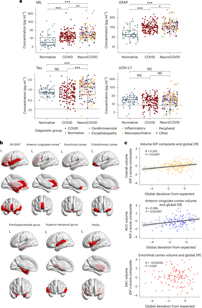
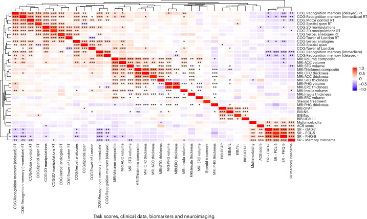
The spectrum, pathophysiology and recovery trajectory of persistent post-COVID-19 cognitive deficits are unknown, limiting our ability to develop prevention and treatment strategies. We report the 1-year cognitive, serum biomarker and neuroimaging findings from a prospective, national study of cognition in 351 COVID-19 patients who required hospitalization, compared with 2,927 normative matched controls. Cognitive deficits were global, associated with elevated brain injury markers and reduced anterior cingulate cortex volume 1 year after COVID-19. Severity of the initial infective insult, postacute psychiatric symptoms and a history of encephalopathy were associated with the greatest deficits. There was strong concordance between subjective and objective cognitive deficits. Longitudinal follow-up in 106 patients demonstrated a trend toward recovery. Together, these findings support the hypothesis that brain injury in moderate to severe COVID-19 may be immune-mediated, and should guide the development of therapeutic strategies.


Conflict of interest statement

Competing interests: R.A.I.B. holds equity and serves as a director for Centile Bioscience Inc. P.J.H. holds equity and serves as director for H2 Cognitive Designs LTD. R.U. was speaker at a promotional educational event for Otuska; and reports consultancy for Vitaris and Springer Healthcare in the past 3 years. T.A.P. reports consultancy for Arialys Therapeutics Inc. and speaker honoraria from Janssen. A.C. is President of the FND Society, Associate Editor of JNNP, and gives independent testimony in court on a range of neuropsychiatric topics. E.B. consults for GSK, SR One, Boehringer Ingelheim and Sosei Heptares. T.S. is Director of The Pandemic Institute, which has received funding from Innova and CSL Seqirus, and Aviva and DAM Health. T.S. was an advisor to the GSK Ebola Vaccine program and the Siemens Diagnostic Program. T.S. chaired the Siemens Healthineers Clinical Advisory Board. T.S. co-chaired the WHO Neuro-COVID task force and sat on the UK Government Advisory Committee on Dangerous Pathogens, and the Medicines and Healthcare Products Regulatory Agency (MHRA) Expert Working Group on COVID-19 vaccines. T.S. advised to the UK COVID-19 Therapeutics Advisory Panel. T.S. was a member of the COVID-19 Vaccines Benefit Risk Expert Working Group for the Commission on Human Medicines committee of the MHRA. T.S. has been a member of the Encephalitis Society since 1998 and President of the Encephalitis Society since 2019. The remaining authors declare no competing interests.

Figures

Fig. 1
Fig. 1. Flow diagram of patients included from the COVID-CNS.
Nationally at least 16,279 patients were screened of whom at least 2,712 were eligible. Matched community data were collected separately and held in a large normative database. ‘Other’ includes autonomic dysfunction, cerebral hypoxic injury, headache, headache and fatigue, hyperkinetic movement disorder, Parkinsonian movement disorder, seizures and speech and sensory. aSix patients with ‘anosmia/ageusia’ reclassified as COVID from NeuroCOVID. The parentheses show n with biomarkers, n with neuroimaging. MOCA, Montreal Cognitive Assessment.
Fig. 2
Fig. 2. Cognitive scores and recovery trajectories.
a, Violin plot of DfE cognition scores by diagnostic group including median (IQR) (black). Statistics compare each group with normative data, n = normative (2,927), cerebrovascular, encephalopathy, inflammatory, neuropsychiatric, peripheral and other. Exact P values are listed in Supplementary Table 8. b, Pattern of deficits in clinical groups by median DfE accuracy and responsive time minus matched community controls across six cognitive tasks. Exact effect sizes and P values in listed Supplementary Table 9. c, Recovery trajectories in NeuroCOVID and COVID patients following postacute assessment. A black dot indicates a single observation, lines connect paired observations between postacute assessment and follow-up 1, and follow-up 1 and follow-up 2. Center line, median; box limits, upper and lower quartiles; whiskers, 1.5× IQR; the dashed line shows normal cognition; the numbers under the x axis show n for each assessment. *P < 0.05, **P < 0.01, ***P < 0.001 two-sided Mann–Whitney U-test, adjusted for multiple comparisons based on the FDR approach in a and b (adjusted for n = 8 and n = 12 comparisons, respectively). 2D, two-dimensional; NS, nonsignificant.
Fig. 3
Fig. 3. Brain injury biomarkers and neuroimaging.
a, Brain injury markers (in pg ml−1) by diagnostic group. Lower limit of quantification (dashed line) if included in scale. Normative values from n = 60 healthy controls. *P < 0.05, **P < 0.01, ***P < 0.001; NS, nonsignificant, unadjusted two-sided Mann–Whitney U. b, Brain regions represented by the IDPs utilized in analyses. These regions are parcellated as per the Desikan-Killiany cortical atlas. For each region and regions combined, IDP composites for thickness and volume were utilized. aIDP composites that have significant correlations with overall cognition (Supplementary Table 2). Created using Matlab and BrainNet Viewer. c, Scatter plots for IDP composite z-scores against GDfE in the overall cohort, using Pearson’s correlation. The black line indicates the line of best fit (least squares method) and gray error band indicates the 95% CI. Significance persisting after adjusted for multiple comparisons based on the FDR approach (adjusted for n = 14 for each IDP composite test). Center line, median; box limits, upper and lower quartiles; whiskers, 1.5× IQR. ACC, anterior cingulate cortex; ERC, entorhinal cortex; UCH-L1, ubiquitin carboxy-terminal hydrolase L1.
Fig. 4
Fig. 4. Heatmap and unsupervised cluster analysis.
Heatmap and unsupervised cluster analysis (Euclidean, complete) in the full cohort (n = 351) of cognitive tasks shaded by correlation (Spearman), including cognition (accuracy and inverse RT), clinical variables, biomarkers and neuroimaging. *P < 0.05, **P < 0.01, ***P < 0.001 two-tailed Spearman correlation adjusted for multiple comparisons (37 ×3 7 matrix, n = 1,369) using the FDR approach. ACB, anticholinergic burden; BIB, brain injury marker; COG, cognitive task; OFC, orbitofrontal cortex; PHG, parahippocampal gyrus; SR, self-report; STG, superior temporal gyrus.

References

    1. Hampshire, A. et al. Multivariate profile and acute-phase correlates of cognitive deficits in a COVID-19 hospitalised cohort. EClinicalMedicine47, 101417 (2022). - PMC - PubMed
    1. Hampshire, A. et al. Cognitive deficits in people who have recovered from COVID-19. EClinicalMedicine39, 101044 (2021). - PMC - PubMed
    1. Heneka, M. T., Golenbock, D., Latz, E., Morgan, D. & Brown, R. Immediate and long-term consequences of COVID-19 infections for the development of neurological disease. Alzheimers Res Ther.12, 69 (2020). - PMC - PubMed
    1. Taquet, M., Geddes, J. R., Husain, M., Luciano, S. & Harrison, P. J. 6-Month neurological and psychiatric outcomes in 236 379 survivors of COVID-19: a retrospective cohort study using electronic health records. Lancet Psychiatry8, 416–427 (2021). - PMC - PubMed
    1. Otani, K., Fukushima, H. & Matsuishi, K. COVID-19 delirium and encephalopathy: pathophysiology assumed in the first 3 years of the ongoing pandemic. Brain Disord.10, 100074 (2023). - PMC - PubMed

MeSH terms

LinkOut - more resources