. 2025 Feb;32(2):315-325.
doi: 10.1038/s41594-024-01397-1. Epub 2024 Sep 23.

Structural insights into CXCR4 modulation and oligomerization

Affiliations

Structural insights into CXCR4 modulation and oligomerization

Kei Saotome et al. Nat Struct Mol Biol. 2025 Feb.

Abstract

Activation of the chemokine receptor CXCR4 by its chemokine ligand CXCL12 regulates diverse cellular processes. Previously reported crystal structures of CXCR4 revealed the architecture of an inactive, homodimeric receptor. However, many structural aspects of CXCR4 remain poorly understood. Here, we use cryo-electron microscopy to investigate various modes of human CXCR4 regulation. CXCL12 activates CXCR4 by inserting its N terminus deep into the CXCR4 orthosteric pocket. The binding of US Food and Drug Administration-approved antagonist AMD3100 is stabilized by electrostatic interactions with acidic residues in the seven-transmembrane-helix bundle. A potent antibody blocker, REGN7663, binds across the extracellular face of CXCR4 and inserts its complementarity-determining region H3 loop into the orthosteric pocket. Trimeric and tetrameric structures of CXCR4 reveal modes of G-protein-coupled receptor oligomerization. We show that CXCR4 adopts distinct subunit conformations in trimeric and tetrameric assemblies, highlighting how oligomerization could allosterically regulate chemokine receptor function.


Conflict of interest statement

Competing interests: All authors are employees of Regeneron Pharmaceuticals and may own stock and/or stock options of the company. W.C.O. is an officer of Regeneron. All funding for this work was provided by Regeneron Pharmaceuticals. The anti-CXCR4 antibody described in this manuscript is a subject of international patent applications (PCT/US2024/044209 and PCT/US2024/044111).

Figures

Fig. 1
Fig. 1. Cryo-EM reconstructions of CXCR4–Gi complexes.
a, Apo CXCR4–Gi complex. b, CXCR4–Gi–CXCL12 complex. Inset, the fit of the CXCL12 N-terminal tail (residues 1–10) in cryo-EM map, represented as a semitransparent surface. The locations of chemokine recognition sites 1 and 2 are labeled. The curved dotted line represents missing density for the distal N terminus of CXCR4, which has been reported to interact with CXCL12. The gray density on the extracellular side may correspond to a partially occupied second protomer of dimeric CXCL12. c, CXCR4–Gi–AMD3100 complex. Inset, the fit of the AMD3100 compound in cryo-EM map.
Fig. 2
Fig. 2. Interactions between CXCR4 and ligands.
a, Expanded view of interaction between the CXCL12 N-terminal tail and CXCR4 orthosteric pocket. Hydrogen-bonding and electrostatic interactions are depicted as dashed lines. b, Expanded view of AMD3100 binding at CXCR4 orthosteric pocket. Asterisks indicate the positions of the two lactam rings, each of which interact with acidic residues. c, Cutaway surface view of CXCR4 orthosteric pocket. The CXCL12 N terminus is shown in stick representation and AMD3100 is shown in sphere representation to illustrate their relative binding positions in the orthosteric pocket.
Fig. 3
Fig. 3. CXCR4 antagonism by REGN7663 monoclonal antibody.
a, CRE luciferase reporter assay showing CXCL12-dependent decrease in signal and block of CXCL12 activity (at 0.5 nM CXCL12) by REGN7663 (light blue). The negative control monoclonal antibody (violet) showed no effect. The IC50 for REGN7663 was calculated to be 2.7 ± 0.1 nM (mean ± s.d.) in antagonist mode from n = 3 independent experiments. RLU, relative luminescence units. b, REGN7663 shows a concentration-dependent increase in signal relative to baseline in the absence of CXCL12, demonstrating inverse agonism. The EC50 for REGN7663 was calculated to be 1.3 ± 0.4 nM in agonist mode (absence of CXCL12) from n = 3 independent experiments. In a,b, representative data from one experiment are shown (the same data for CXCL12 are shown as solid black circles in a,b to allow a comparison to monoclonal antibody data). c, Cryo-EM reconstruction of CXCR4EM–Gi–REGN7663 Fab complex, with each polypeptide chain colored differently. d, Top-down view of CXCR4 (yellow) with CDR loops of bound REGN7663 shown (blue, heavy chain (HC); cyan, light chain (LC)). e, Electrostatic interaction between CDR-H3 of REGN7663 and CXCR4 orthosteric pocket-facing residue E288. Source data
Fig. 4
Fig. 4. Inactive CXCR4 structure and structural basis of activation.
a, Cryo-EM reconstruction of inactive CXCR4EM–REGN7663 Fab complex (CXCR4, pink; REGN7663 heavy chain, gray; REGN7663 light chain, white). b, Structural alignment of inactive CXCR4 (pink) and active CXCR4 (yellow); the CXCR4EM–Gi–REGN7663 Fab complex was used for alignment. Left, side view; right, bottom-up view. The green block arrows depict conformational transitions from inactive to active CXCR4. c, Expanded view showing CXCL12 N terminus (cyan) binding to active CXCR4 (yellow). Inactive CXCR4 (pink) is shown for comparison and residues important for transmitting chemokine binding into activation are shown in stick representation. d, Expanded view of Gαi (light green) binding to active CXCR4 (yellow). Residues participating in the interaction are shown in stick representation and labeled (Gαi residue labels are underlined). Electrostatic interactions are highlighted with dashed lines.
Fig. 5
Fig. 5. Oligomeric CXCR4 structures.
a, Cryo-EM reconstruction of CXCR4 trimer in complex with REGN7663 Fab. b,c, Side (b) and top-down (c) views of CXCR4 trimer structure. TM helices are shown in cylinder representation and bound lipids are shown in stick representation. Fab molecules are omitted for clarity. d, Cryo-EM reconstruction of CXCR4 tetramer in complex with REGN7663 Fab. e,f, Side (e) and top-down (f) views of CXCR4 tetramer structure. g, Side (left) and top (right) views of previously reported dimeric crystal structure of CXCR4. h, Top-down view of a CXCR4 protomer (gray) showing the positions of neighboring subunits from a dimer (orange), trimer (cyan) and tetramer (magenta).
Fig. 6
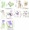
Fig. 6. Oligomeric interfaces and protomer conformations.
a, Interprotomer interface of CXCR4 trimer. Interface residues are shown in stick representation and labeled. b, Interprotomer interface of CXCR4 tetramer. Interface residues and modeled cholesterol are shown in stick representation. The density corresponding to cholesterol is shown as a transparent gray surface. c, Bottom-up view showing position of cholesterol at the tetramer interface. d, Structural alignment of TM6 and TM1 at the trimer (gray) and tetramer (blue and magenta, with cholesterol (yellow) shown in stick representation). e,f, Side (e) and bottom-up (f) views of protomeric structures of trimeric (cyan) and tetrameric (magenta) CXCR4. Binding of the Gαi α5 helix (gray) is prevented by steric clash. g, Structural alignment of trimeric CXCR4 protomer (cyan) and active CXCR4 protomer (yellow). Red asterisks highlight the distinct positions of ICL3 and TM7. h, Structural alignment of tetrameric CXCR4 protomer (magenta) and inactive CXCR4 (pink).
Extended Data Fig. 1
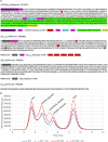
Extended Data Fig. 1. Protein constructs and FSEC-based protein screening.
a, Primary structures of protein constructs used in structural studies. b, Fluorescence-detection size exclusion chromatography (FSEC) screening of wild type CXCR4 (blue) and N119S-containing CXCR4 (referred to as CXCR4EM, red) in the presence (solid lines) or absence (dashed lines) of added Gi. The nominally wild type CXCR4 construct was identical to that shown in (a) without the N119S point mutation. Chromatograms are annotated with presumed peak positions of various species present in the samples. Source data
Extended Data Fig. 2
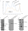
Extended Data Fig. 2. Purification of CXCR4 complexes.
a, Size-exclusion chromatography (SEC) traces for a CXCR4EM purification with added Gi (blue), and for a purification prepared in the absence of exogenously added Gi (orange). CXCR4EM/Gi complex chromatogram is representative of four purifications performed each yielding similar results, whereas purification of CXCR4EM in the absence of added Gi was conducted once. b,c SDS-PAGE (4–20% Tris-Glycine) showing the subunit content and purity of prepared cryoEM samples for CXCR4EM/Gi complex (b, representative of four preparations yielding similar results) and CXCR4EM prepared in the absence of added Gi (c, prepared once). 2% (v/v) 2-Mercaptoetanol was present in the SDS-PAGE samples prior to loading. d, Blue native (BN) PAGE (4–16% Bis-Tris) of SEC-purified CXCR4EM monomer and oligomer samples prepared in the absence of added Gi. BN PAGE analysis was conducted on one sample. Source data
Extended Data Fig. 3
Extended Data Fig. 3. CryoEM reconstruction of Apo CXCR4EM/Gi, CXCL12-bound CXCR4EM/Gi, AMD3100-bound CXCR4EM/Gi.
a-d, FSC curve (a), particle angular distribution plot (b), local resolution map calculated in cryoSPARC (c), and map/model fits of selected regions (d) for Apo CXCR4EM/Gi. e-h, FSC curve (e), particle angular distribution plot (f), local resolution map calculated in cryoSPARC (g), and map/model fits of selected regions (h) for CXCL12-bound CXCR4EM/Gi. i, two views showing putative fit of a CXCL12 dimer (arranged on the basis of PDB 3GV3) into cryoEM map, shown at 4 sigma in PyMOL. j-m, FSC curve (j), particle angular distribution plot (k), local resolution map calculated in cryoSPARC (l), and map/model fits of selected regions (m) for AMD3100-bound CXCR4EM/Gi.
Extended Data Fig. 4
Extended Data Fig. 4. Structural comparisons of chemokine receptor structures.
a, structural alignment of active CXCR4 (yellow, this study, CXCL12/Gi-bound complex) and inactive, antagonist-bound CXCR4 (cyan, PDB 3OE0). Magenta block arrows depict movement of TM6. b, Alignment of CXCR4/CXCL12 complex (yellow) with CXCR2/CXCL8 complex (brown, PDB 6LFO), and CCR2/CCL2 complex (pink, PDB 7XA3). G protein models are omitted for clarity. c, Receptor-based alignment of CXCR4/CXCL12 (yellow/pink) with ACKR3/CXCL12 (gray/black, PDB 7SK3). Arrow highlights different docking orientations of CXCL12 onto the two receptors. d, expanded views of showing different binding modes of CXCL12 N-termini (pink in CXCR4 complex and black in ACKR3 complex) in CXCR4 and ACKR3. e, alignment of CXCR4/CXCL12 complex (yellow/pink) and CXCR4/vMIP-II (green/blue). Inset shows expanded views of chemokine N-terminal positions within orthosteric pocket, highlighting the positions of toggle switch residue W252 in sticks.
Extended Data Fig. 5
Extended Data Fig. 5. CryoEM reconstruction of REGN7663 Fab/CXCR4EM/Gi and REGN7663 Fab/CXCR4EM.
a-d, FSC curve (a), particle angular distribution plot (b), local resolution map calculated in cryoSPARC (c), and map/model fits of selected regions (d) for REGN7663 Fab/CXCR4EM/Gi. e, f, Expanded view of contacts between REGN7663 Fab (light chain in cyan, heavy chain in blue) and CXCR4 ECL2 (e) and N-term (f). Epitope and paratope residues are shown as sticks and labeled, and apparent salt bridges/hydrogen bonds between mAb and receptor are shown as dashed lines. g, structural alignment of CXCR4 bound to CXCL12 and REGN7663 Fab. N-term. and ECL2 are colored green (CXCL12-bound) or magenta (REGN7663 Fab-bound) to highlight their different positions. h-k, FSC curve (h), particle angular distribution plot (i), local resolution map calculated in cryoSPARC (j), and map/model fits of TM helices (k) for REGN7663 Fab/CXCR4EM without Gi. l, aligned structures of CXCR4/REGN7663 Fab complex in the inactive (pink) and active (yellow, Gi-bound) conformations. Note the REGN7663 Fab variable region and cytoplasmic half of the CXCR4 domain are mostly superimposable.
Extended Data Fig. 6
Extended Data Fig. 6. Structural features of Gi-bound chemokine receptors.
a, alignment of CXCR4/Gi complexes bound to CXCL12 (yellow), REGN7663 Fab (magenta), AMD3100 (green) or in the absence of ligand (apo, gray). Inset shows expanded view around E288 residue. Bound CXCL12 is shown as yellow transparent surface and sticks, highlighting how it enforces a rotameric change of E288 and slight shift of TM7 in the CXCL12-bound complex. b, receptor-based alignment showing architecture of various chemokine/chemokine receptor/Gi complexes. c, expanded view showing docking of Gαi α5 helix into cytoplasmic pocket of chemokine receptors. Note that the Gαi α5 helix is positioned closer to ICL2 in CC chemokine receptor complexes, while it is closer to ICL3 in CXC chemokine receptor complexes.
Extended Data Fig. 7
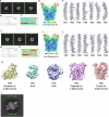
Extended Data Fig. 7. CryoEM of CXCR4 oligomers.
a-e, example 2D class averages obtained from tilted data collection (a), FSC curves (b), angular distribution plot (c), local resolution map (d) and model/map fits of TM helices (e) of trimeric CXCR4EM/REGN7663 Fab complex. f-j, example 2D class averages obtained from tilted data collection (f), FSC curves (g), angular distribution plot (h), local resolution map (i) and model/map fits of TM helices (j) of tetrameric CXCR4EM/REGN7663 Fab complex. k, Output maps from ab initio reconstruction conducted on the oligomeric CXCR4EM/REGN7663 Fab particles. Particles belonging to classes of tetramer with 4 fabs bound or trimer with 3 fabs bound were selected for further processing. l, An example 2D class average showing an anti-parallel dimer of CXCR4EM/Gi.
Extended Data Fig. 8
Extended Data Fig. 8. Lipids resolved in oligomeric structures of CXCR4.
a-c, side (a), top-down (b) and bottom-up (c) views of trimeric CXCR4, highlighting positions of built lipid molecules. Cholesterols (chol.) are shown as yellow sticks and phosphatidic acid (PA) are shown as green sticks. d, fit of lipid molecules (shown as sticks) to map (transparent blue surface) in trimeric CXCR4. Chol. 1 and chol 2. refer to lipids labeled in a-c. e-g, side (e), top-down (f) and bottom-up (g) views of tetrameric CXCR4, highlighting positions of built lipid molecules. h, fit of lipid molecules (shown as sticks) to map (transparent blue surface) in tetrameric CXCR4.
Extended Data Fig. 9
Extended Data Fig. 9. Structural analysis of dimeric and oligomeric structures of CXCR4.
a,b, top-down view of hypothetical models of four CXCR4 trimers (a) or five CXCR4 tetramers (b) clustered via dimeric interfaces (red ovals) observed in crystal structures. c,d, a single subunit from dimeric x-ray structure of CXCR4 (receptor in yellow, ICL3-fused T4 lysozyme (T4L) in gray) aligned to trimeric CXCR4 (c) or tetrameric CXCR4 (d). Side and top views are shown. Red symbols indicate steric clash between T4L and neighboring protomers that would prevent trimer or tetramer assembly. The steric hindrance caused by fused T4L may explain why dimeric CXCR4 was favored over trimeric or tetrameric CXCR4 in previous crystallographic studies.
Extended Data Fig. 10
Extended Data Fig. 10. FSEC analysis of CXCR4 oligomerization interface mutants.
a, FSEC chromatograms of control construct CXCR4EM (black trace) and its mutants (colored traces) tracking GFP fluorescence. The same chromatogram for the control are shown in each for comparison. Gray shaded regions indicate elution times corresponding to CXCR4 oligomer (7.8-8.8 min) and monomer (9.6-10 min). b, ratio of oligomer to monomer peak areas, calculated according to shaded regions in a. horizontal dotted line corresponds to mean value for CXCR4EM. Column heights and error bars show mean and standard deviations calculated from n = 3 (mutant constructs) or n = 4 (CXCR4EM control) FSEC experiments using two independently generated baculoviruses for each construct. Note that several mutants (I257W, I300W, F276A/W283A, F276A, W283A) showed poor chromatographic behavior overall, presumably due to poor expression or stability in detergent. Source data

Similar articles

Cited by

References

    1. Hughes, C. E. & Nibbs, R. J. B. A guide to chemokines and their receptors. FEBS J.285, 2944–2971 (2018). - PMC - PubMed
    1. Busillo, J. M. & Benovic, J. L. Regulation of CXCR4 signaling. Biochim. Biophys. Acta1768, 952–963 (2007). - PMC - PubMed
    1. Britton, C., Poznansky, M. C. & Reeves, P. Polyfunctionality of the CXCR4/CXCL12 axis in health and disease: implications for therapeutic interventions in cancer and immune-mediated diseases. FASEB J.35, e21260 (2021). - PubMed
    1. Guo, F. et al. CXCL12/CXCR4: a symbiotic bridge linking cancer cells and their stromal neighbors in oncogenic communication networks. Oncogene35, 816–826 (2016). - PubMed
    1. Feng, Y., Broder, C. C., Kennedy, P. E. & Berger, E. A. HIV-1 entry cofactor: functional cDNA cloning of a seven-transmembrane, G protein-coupled receptor. Science272, 872–877 (1996). - PubMed

LinkOut - more resources