

The role of cell-cell and cell-matrix junctional complexes in sebaceous gland homeostasis and differentiation
- PMID: 39313816
- PMCID: PMC11421122
- DOI: 10.1186/s12964-024-01835-z
The role of cell-cell and cell-matrix junctional complexes in sebaceous gland homeostasis and differentiation
Abstract
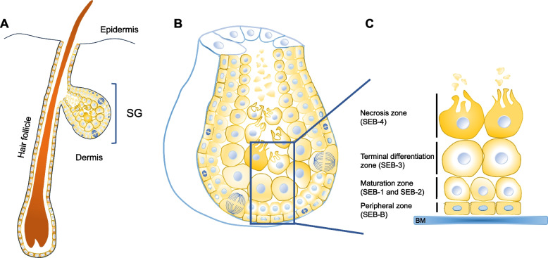
Sebaceous glands (SG) are essential for maintaining skin integrity, as their lipid-rich secretion (sebum) lubricates and protects the epidermis and hairs. In addition, these glands have an emerging role in immunomodulation and may affect whole-body energy metabolism, besides being an appealing model for research in topics as lipogenesis, stem cell biology and tumorigenesis. In spite of the increasing interest in studying SGs pathophysiology, sebocyte cell-cell and cell-matrix adhesion processes have been only superficially examined, and never in a systematic way. This is regrettable considering the key role of cellular adhesion in general, the specific expression pattern of indivdual junctional complexes, and the reports of structural changes in SGs after altered expression of adhesion-relevant proteins. Here, we review the available information on structural and functional aspects of cell-cell and cell-matrix junctions in sebocytes, and how these processes change under pathological conditions. This information will contribute for better understanding sebocyte differentiation and sebum secretion, and may provide hints for novel therapeutic strategies for skin diseases.
Keywords: Cell–cell adhesion; Cell–matrix adhesion; Junctional complexes; Sebaceous glands; Sebum.
© 2024. The Author(s).
Conflict of interest statement
The authors declare no competing interests.
Figures


Similar articles
-
Neuroendocrinology and neurobiology of sebaceous glands.Biol Rev Camb Philos Soc. 2020 Jun;95(3):592-624. doi: 10.1111/brv.12579. Epub 2020 Jan 22. Biol Rev Camb Philos Soc. 2020. PMID: 31970855 Review.
-
Sebaceous gland: Milestones of 30-year modelling research dedicated to the "brain of the skin".Exp Dermatol. 2020 Nov;29(11):1069-1079. doi: 10.1111/exd.14184. Epub 2020 Sep 25. Exp Dermatol. 2020. PMID: 32875660
-
A spatial portrait of the human sebaceous gland transcriptional program.J Biol Chem. 2024 Jul;300(7):107442. doi: 10.1016/j.jbc.2024.107442. Epub 2024 Jun 3. J Biol Chem. 2024. PMID: 38838779 Free PMC article.
-
TGFβ signaling regulates lipogenesis in human sebaceous glands cells.BMC Dermatol. 2013 Jan 23;13:2. doi: 10.1186/1471-5945-13-2. BMC Dermatol. 2013. PMID: 23343495 Free PMC article.
-
Primary sebocytes and sebaceous gland cell lines for studying sebaceous lipogenesis and sebaceous gland diseases.Exp Dermatol. 2018 May;27(5):484-488. doi: 10.1111/exd.13513. Epub 2018 Apr 15. Exp Dermatol. 2018. PMID: 29451719 Review.
Cited by
-
The metabolic underpinnings of sebaceous lipogenesis.Commun Biol. 2025 Apr 27;8(1):670. doi: 10.1038/s42003-025-08105-9. Commun Biol. 2025. PMID: 40289206 Free PMC article. Review.
-
The Sebaceous Gland: A Key Player in the Balance Between Homeostasis and Inflammatory Skin Diseases.Cells. 2025 May 20;14(10):747. doi: 10.3390/cells14100747. Cells. 2025. PMID: 40422250 Free PMC article. Review.
References
-
- Smith KR, Thiboutot DM. Thematic review series: skin lipids. Sebaceous gland lipids: friend or foe? J Lipid Res. 2008;49:271–81. - PubMed
-
- Schneider MR, Paus R. Sebocytes, multifaceted epithelial cells: lipid production and holocrine secretion. Int J Biochem Cell Biol. 2010;42:181–5. - PubMed
-
- Nicolaides N. Skin lipids: their biochemical uniqueness. Science. 1974;186:19–26. - PubMed
Publication types
MeSH terms
LinkOut - more resources
Full Text Sources
Molecular Biology Databases
