. 2024 Sep 23;55(1):114.
doi: 10.1186/s13567-024-01363-1.

The immunoglobulin M-degrading enzyme of Streptococcus suis (IdeSsuis) leads to long-lasting inhibition of the activation of porcine IgM-secreting B cells

Affiliations

The immunoglobulin M-degrading enzyme of Streptococcus suis (IdeSsuis) leads to long-lasting inhibition of the activation of porcine IgM-secreting B cells

Annika Katharina Breitfelder et al. Vet Res. .

Abstract

Streptococcus suis (S. suis) is one of the most important porcine pathogens, causing severe pathologies such as meningitis or polyarthritis. It is also a very successful colonizer of mucosal surfaces. The IgM-degrading enzyme of S. suis (IdeSsuis) specifically cleaves porcine IgM, which results in complement evasion. On the basis of our previous finding that IdeSsuis also cleaves the IgM B cell receptor in vitro, we verified IgM B cell receptor cleavage ex vivo in whole regional lymph nodes and investigated the working hypothesis that this IgM B cell receptor cleavage results in a long-lasting impaired B cell function. The number of IgM-secreting cells was determined via ELISpot analysis after porcine peripheral blood mononuclear cells had initially been treated with different recombinant S. suis proteins and subsequently stimulated with interleukin-2 and the toll-like receptor 7/8 ligand R848. Compared with treatment with medium or recombinant muramidase-released protein, treatment with rIdeSsuis but also with a cleavage-deficient variant led to a reduction in the number of IgM-secreting cells as well as the level of secreted IgM. Flow cytometry analysis confirmed that the IgM B cell receptor was cleaved only by rIdeSsuis, and the receptor recovered to pretreatment levels on day 2 after treatment. Flow cytometry analysis of B and T cells incubated with fluorescein-labelled recombinant proteins revealed that different rIdeSsuis variants bind specifically to B cells, most prominently the cleavage-deficient variant. Our results indicate that in vitro interference of rIdeSsuis with the IgM B cell receptor results in long-lasting impaired IgM secretion by B cells after toll-like receptor activation. Further studies are warranted to prove that the modulation of B cell function by IdeSsuis could play a role in vivo.

Keywords: Streptococcus suis; B cell receptor; B cell receptor cleavage; ELISpot; IdeSsuis; IgM.


Conflict of interest statement

The authors declare that they have no competing interests.

Figures

Figure 1
Figure 1
The IgM BCR is cleaved by rIdeSsuis_h in local lymph nodes ex vivo. Regional lymph nodes (Ln. cervicalis superficialis dorsalis, Ln. subiliacus, Ln. inguinalis superficialis) were injected with rIdeSsuis_h or rIdeSsuis_h_C195S ex vivo, and the percentage of IgM Fc+ F(abˈ)2+ B cells among the isolated cells was investigated via flow cytometry.
Figure 2
Figure 2
ELISpot procedure to analyze the number of IgM-secreting cells. Porcine PBMC were incubated in cell culture medium alone or with (i) rIdeSsuis_homologue (rIdeSsuis_h), (ii) rIdeSsuis_homologue_C195S (rIdeSsuis_h_C195S), (iii) rIdeSsuis_C_domain, or (iv) rMRP for 45 min before protein removal by washing. Treatment with these recombinant proteins was performed before (A) or after (B) 3 d of incubation in stimulation medium (supplemented with IL-2 and R848). After protein treatment and subsequent stimulation, the culture supernatant was collected for IgM ELISA, and the cells that were not used for ELISpot analysis were analysed via flow cytometry. The cells were washed and readjusted before being seeded in the ELISpot plate at 1 × 104 cells/well. The wells were coated with an anti-IgM antibody to capture secreted IgM. After 24 h, the cells were removed, and the captured IgM was detected with a second, biotinylated anti-IgM antibody. Streptavidin transforms the added substrate to a purple color, which forms visible spots exactly at the place where an IgM-secreting cell has been. This figure was created with Biorender.
Figure 3
Figure 3
ELISpot and ELISA analysis of porcine cells after treatment with different recombinant proteins. A, B Incubation with rIdeSsuis_homologue and rIdeSsuis_homologue_C195S before stimulation for 3 days reduced the number of IgM-secreting cells. Porcine PBMC were incubated in cell culture medium alone or with (i) rIdeSsuis_homologue (rIdeSsuis_h), (ii) rIdeSsuis_homologue_C195S (rIdeSsuis_h_C195S), (iii) rIdeSsuis_C_domain or (iv) rMRP (all 4 µg/106 cells, 45 min) before protein removal by washing and subsequent incubation in stimulation medium (supplemented with IL-2 and R848) for 3 days. A Exemplary ELISpot wells. B ELISpot analysis of porcine PBMC (n = 14). Statistical analysis was conducted with ordinary one-way ANOVA with Tukey’s multiple comparisons test (p < 0.001 ***, p < 0.01 **, p < 0.05 *, p > 0.05 ns). Only comparisons with p < 0.05 are shown. The bars and error bars represent the means and standard deviations, respectively. C Incubation with rIdeSsuis_homologue and rIdeSsuis_homologue_C195S before stimulation for 3 days also reduced the level of secreted IgM. IgM ELISA of ELISpot culture supernatants collected after 3 days of incubation in stimulation medium. Stimulation medium without PBMC or supernatant from unstimulated cells served as a negative control. Statistical analysis was conducted with the Friedmann test with Dunn’s multiple comparisons test (p < 0.05 *, p > 0.05 ns). Only comparisons with p < 0.05 are shown. The bars represent the medians. D Incubation with recombinant proteins after 3 days of stimulation did not impair the number of IgM-secreting cells. ELISpot analysis of porcine PBMC (n = 7). The cells were incubated in stimulation medium for 3 days before being washed and incubated in cell culture medium alone or with different recombinant proteins (as described above). Statistical analysis was conducted with ordinary one-way ANOVA and Tukey’s multiple comparisons test (p > 0.05 ns). Only comparisons with p < 0.05 are shown. The bars and error bars represent the means and standard deviations, respectively. Note: Symbols represent different treatments, and colours represent individual pigs.
Figure 4
Figure 4
Expression of the IgM BCR on PBMC reaches levels comparable to those of cells treated with medium or other recombinant proteins two days after treatment with the rIdeSsuis_homologue. Porcine PBMC were incubated in cell culture medium alone or with (i) rIdeSsuis_homologue (rIdeSsuis_h), (ii) rIdeSsuis_homologue_C195S (rIdeSsuis_h_C195S), (iii) rIdeSsuis_C_domain or (iv) rMRP (all 4 µg/106 cells, 45 min) before protein removal by washing. Samples were taken immediately after washing (0 h) or after 2 days or 3 days of incubation in stimulation medium (supplemented with IL-2 and R848). The cells were stained and analysed for the percentage of IgM Fc+ F(abˈ)2+ B cells (A) and the respective median fluorescence intensity (MFI, B).
Figure 5
Figure 5
Incubation with recombinant proteins does not impair cell viability. Porcine PBMC (n = 3, representative data points) were incubated in cell culture medium alone or with (i) rIdeSsuis_homologue (rIdeSsuis_h), (ii) rIdeSsuis_homologue_C195S (rIdeSsuis_h_C195S), (iii) rIdeSsuis_C-domain or iiii) rMRP (all 4 µg/106 cells, 45 min) before protein removal by washing. Samples were taken immediately after washing (0 h) or after 2 days or 3 days of incubation in stimulation medium (supplemented with IL-2 and R848). The cells were stained with live‒dead stain and analysed for viable cells within single lymphocytes via flow cytometry.
Figure 6
Figure 6
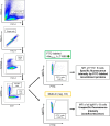
Incubation with FITC-labelled recombinant proteins: Gating strategy. Representative pseudocolor plots are shown. Porcine PBMC were incubated in cell culture medium alone (negative control) or with FITC-labelled recombinant proteins (4 µg/106 cells, 45 min). Viable cells were gated for the B-cell marker CD79α and surface IgM. Finally, the percentage of FITC+ IgM + B cells was analysed. The median fluorescence intensity (MFI) of IgM+ B cells incubated in medium alone was considered autofluorescence. The MFI of the FITC+ IgM+ B cells was considered to be specific because they were incubated with the FITC-labelled proteins.
Figure 7
Figure 7
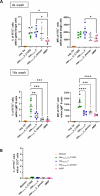
Cleavage-deficient point-mutant rIdeSsuis_homologue_C195S binds more strongly to IgM+ B cells than does rIdeSsuis_homologue. A Flow cytometry analysis of FITC+ cells. Porcine PBMC (n = 6) were incubated with FITC-labelled proteins: (i) rIdeSsuis_homologue (rIdeSsuis_h), (ii) rIdeSsuis_homologue_C195S (rIdeSsuis_h_C195S), (iii) rIdeSsuis_C_domain or (iv) rMRP (all 4 µg/106 cells, 45 min). Upper row: Cells were analyzed directly after fixation, with a total of 4 washing steps before analysis. Lower row: Cells were stained for IgM+ B cells for a total of 14 washing steps before analysis. The percentage of FITC+ cells (left hand side) and the respective median fluorescence intensity (MFI, right hand side) are shown. Statistical analysis was conducted with ordinary one-way ANOVA with Tukey’s multiple comparisons test (p < 0.0001 ****, p < 0.001 ***, p < 0.01 **, p < 0.05 *, p > 0.05 ns). Only comparisons with p < 0.05 are shown. The bars and error bars represent the means and standard deviations, respectively. B FITC-labelled recombinant proteins do not bind to CD3+ T cells. Porcine PBMC (n = 6) were incubated in cell culture medium alone or with the indicated recombinant proteins as described above. The CD3 + T cells were stained and analysed by flow cytometry (for a total of 14 washing steps before analysis).

Similar articles

References

    1. Gottschalk M, Segura M (2019) Streptococcosis. In: Zimmerman JJ, Karriker LA, Ramirez A, Schwartz KJ, Stevenson GW, Zhang J (eds) Disease of swine, 11th edn. Wiley-Blackwell, Hoboken NJ, pp 934–950
    1. Baele M, Chiers K, Devriese LA, Smith HE, Wisselink HJ, Vaneechoutte M, Haesebrouck F (2001) The gram-positive tonsillar and nasal flora of piglets before and after weaning. J Appl Microbiol 91:997–1003 - PubMed
    1. Torremorell M, Calsamiglia M, Pijoan C (1997) Colonization of suckling pigs by Streptococcus suis with particular reference to pathogenic serotype 2 strains. Can J Vet Res 62:21–26 - PMC - PubMed
    1. Gajdács M, Németh A, Knausz M, Barrak I, Stájer A, Mestyán G, Melegh S, Nyul A, Tóth Á, Ágoston Z, Urbán E (2020) Streptococcus suis: an underestimated emerging pathogen in Hungary? Microorganisms 8:1292 - PMC - PubMed
    1. Wertheim HFL, Nghia HDT, Taylor W, Schultsz C (2009) Streptococcus suis: an emerging human pathogen. Clin Infect Dis 48:617–625 - PubMed

MeSH terms

LinkOut - more resources